
















Copyright: ©Author(s) 2026.
World J Gastrointest Oncol. Nov 15, 2026; 18(11): 124574
Published online Nov 15, 2026. doi: 10.4251/wjgo.124574
Published online Nov 15, 2026. doi: 10.4251/wjgo.124574
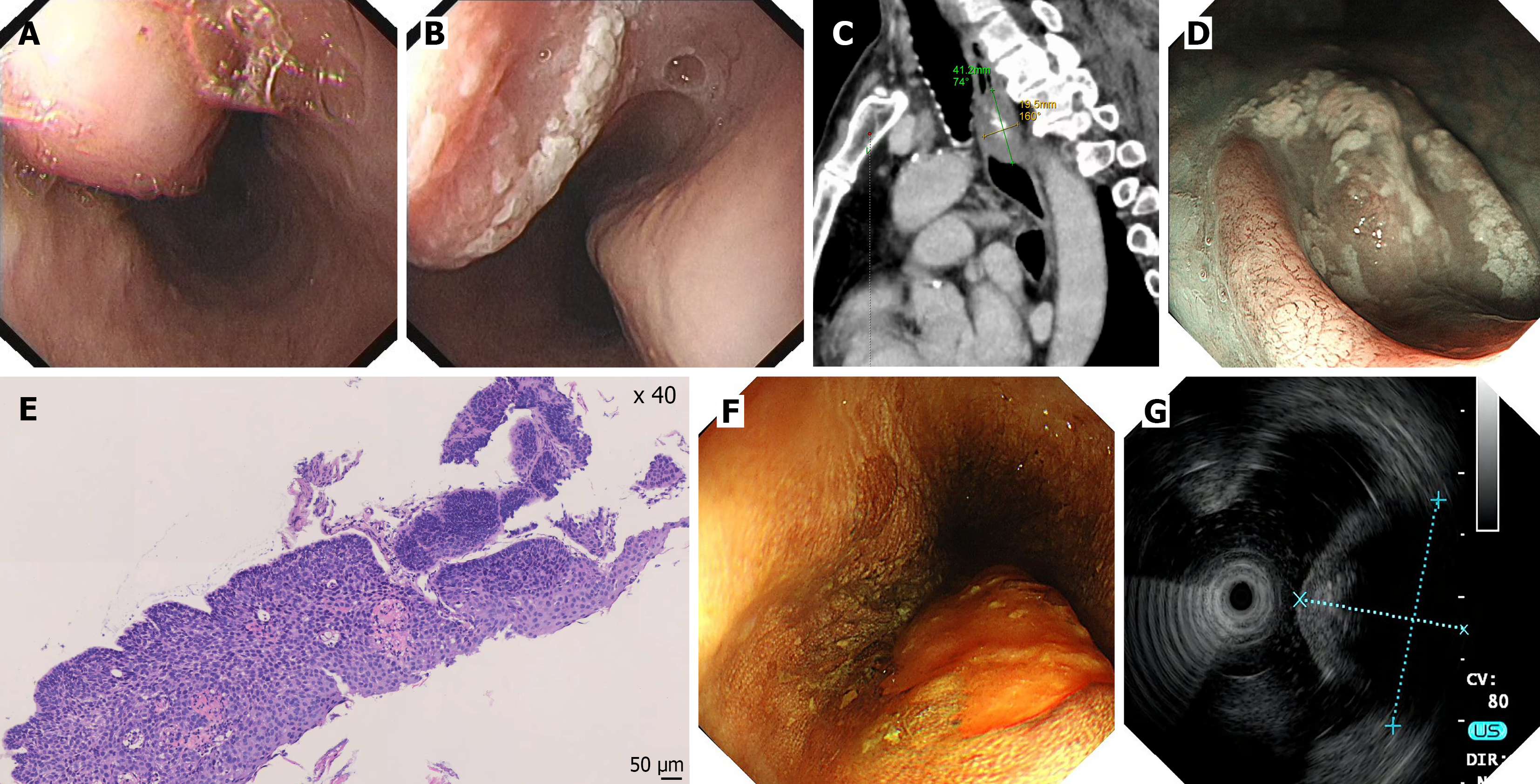
Figure 1 Preoperative multimodal evaluation of the esophageal lesions.
A: White-light endoscopy showing a submucosal tumor-like elevation in the upper esophagus; B: White-light endoscopy showing a superficially elevated mucosal lesion with grayish-white exudate on the surface; C: Contrast-enhanced chest computed tomography of the chest (sagittal view) shows calcification within the lesion; D: Magnifying narrow-band imaging endoscopy reveals type B2 intrapapillary capillary loops; E: Preoperative biopsy histopathology showing high-grade intraepithelial neoplasia of the esophageal squamous epithelium; F: Lugol iodine staining showing an unstained area of the lesion; G: Endoscopic ultrasonography shows the lesion as a hypoechoic mass with local hyperechoic areas.
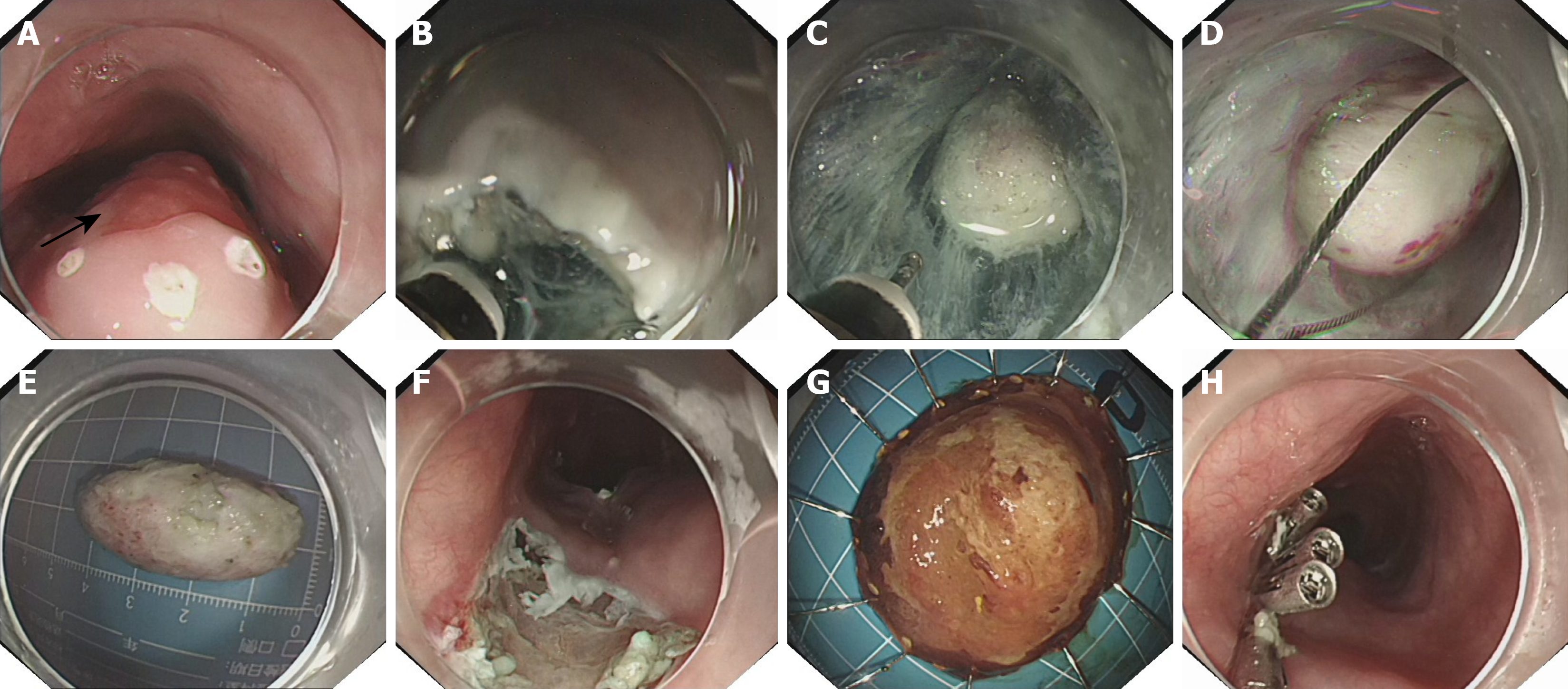
Figure 2 Endoscopic resection procedure for the esophageal submucosal tumor and adjacent mucosal lesion.
A: Esophageal calcifying fibrous tumor (CFT) (21-24 cm, submucosal elevation) and squamous cell carcinoma (ESCC) (24-26 cm, reddish mucosal lesion, at the black arrow position); B: Establishment of a submucosal tunnel (approximately 3 cm oral to the CFT, along the submucosal layer toward the anal side); C: Creation of the submucosal tunnel during submucosal tunneling endoscopic resection, with adequate dissection of the tissue surrounding the tumor; D: Retrieval of the tumor with a snare; E: Gross appearance of the resected submucosal tumor specimen; F: Post-endoscopic submucosal dissection (ESD) wound after resection of the mucosal lesion; G: Resected mucosal lesion specimen (ESCC) after ESD; H: Closure of the tunnel entry and the ESD-induced mucosal defect with clips.
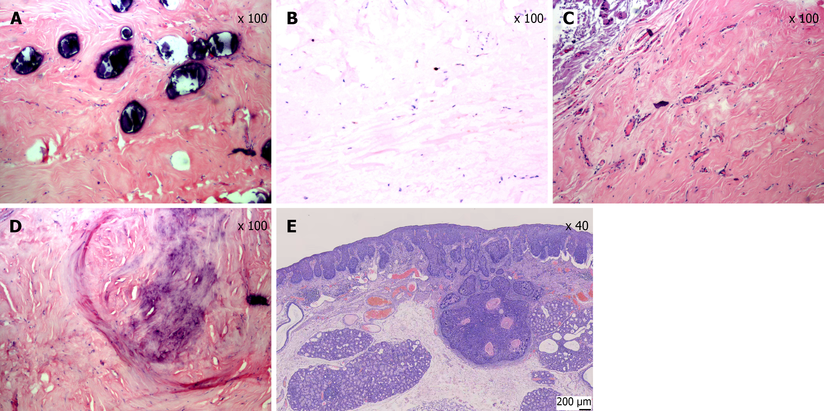
Figure 3 Histopathological and immunohistochemical features of the esophageal calcifying fibrous tumor and squamous cell carcinoma.
A: High-power hematoxylin and eosin (HE) staining (× 100) showing multiple basophilic calcified deposits within dense collagenous stroma. Scale bar = 100 μm; B: Immunohistochemical staining for Ki-67 (× 100) showing a low proliferation index (< 1%), consistent with a benign lesion. Scale bar = 100 μm; C: Low-power HE staining (× 100) showing paucicellular dense collagenous tissue with scattered bland spindle cells, without significant cytologic atypia or increased mitotic activity. Scale bar = 100 μm; D: High-power HE staining (× 100) showing chondroid-like hyalinized stroma with focal calcification. Scale bar = 100 μm; E: HE staining (× 40) of the mucosal lesion showing moderately differentiated esophageal squamous cell carcinoma invading the submucosa. Scale bar = 200 μm.
- Citation: Zhou Y, Duan H, Chen ZY, He DX. Endoscopic treatment of calcifying fibrous tumor of the esophagus combined with esophageal cancer: A case report. World J Gastrointest Oncol 2026; 18(11): 124574
- URL: https://www.wjgnet.com/1948-5204/full/v18/i11/124574.htm
- DOI: https://dx.doi.org/10.4251/wjgo.124574