
















Copyright: ©Author(s) 2026.
World J Gastrointest Oncol. Nov 15, 2026; 18(11): 124574
Published online Nov 15, 2026. doi: 10.4251/wjgo.124574
Published online Nov 15, 2026. doi: 10.4251/wjgo.124574
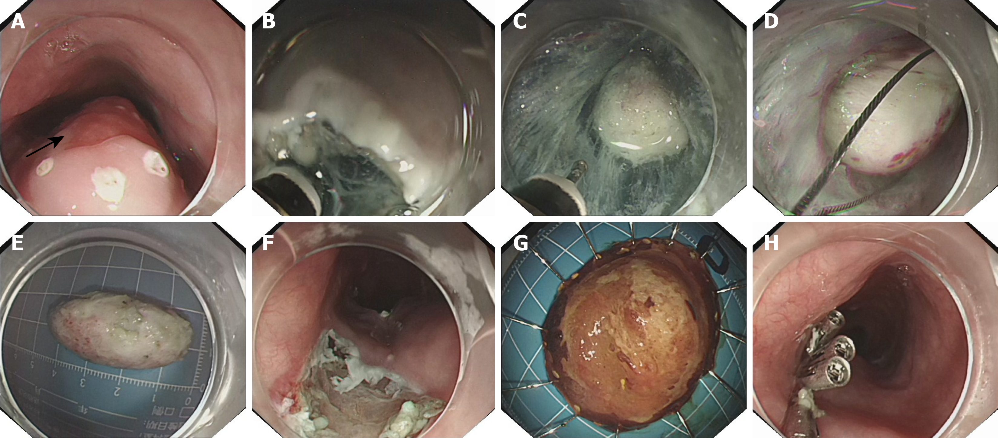
Figure 2 Endoscopic resection procedure for the esophageal submucosal tumor and adjacent mucosal lesion.
A: Esophageal calcifying fibrous tumor (CFT) (21-24 cm, submucosal elevation) and squamous cell carcinoma (ESCC) (24-26 cm, reddish mucosal lesion, at the black arrow position); B: Establishment of a submucosal tunnel (approximately 3 cm oral to the CFT, along the submucosal layer toward the anal side); C: Creation of the submucosal tunnel during submucosal tunneling endoscopic resection, with adequate dissection of the tissue surrounding the tumor; D: Retrieval of the tumor with a snare; E: Gross appearance of the resected submucosal tumor specimen; F: Post-endoscopic submucosal dissection (ESD) wound after resection of the mucosal lesion; G: Resected mucosal lesion specimen (ESCC) after ESD; H: Closure of the tunnel entry and the ESD-induced mucosal defect with clips.
- Citation: Zhou Y, Duan H, Chen ZY, He DX. Endoscopic treatment of calcifying fibrous tumor of the esophagus combined with esophageal cancer: A case report. World J Gastrointest Oncol 2026; 18(11): 124574
- URL: https://www.wjgnet.com/1948-5204/full/v18/i11/124574.htm
- DOI: https://dx.doi.org/10.4251/wjgo.124574