
















Copyright: ©Author(s) 2026.
World J Gastrointest Oncol. Nov 15, 2026; 18(11): 124574
Published online Nov 15, 2026. doi: 10.4251/wjgo.124574
Published online Nov 15, 2026. doi: 10.4251/wjgo.124574
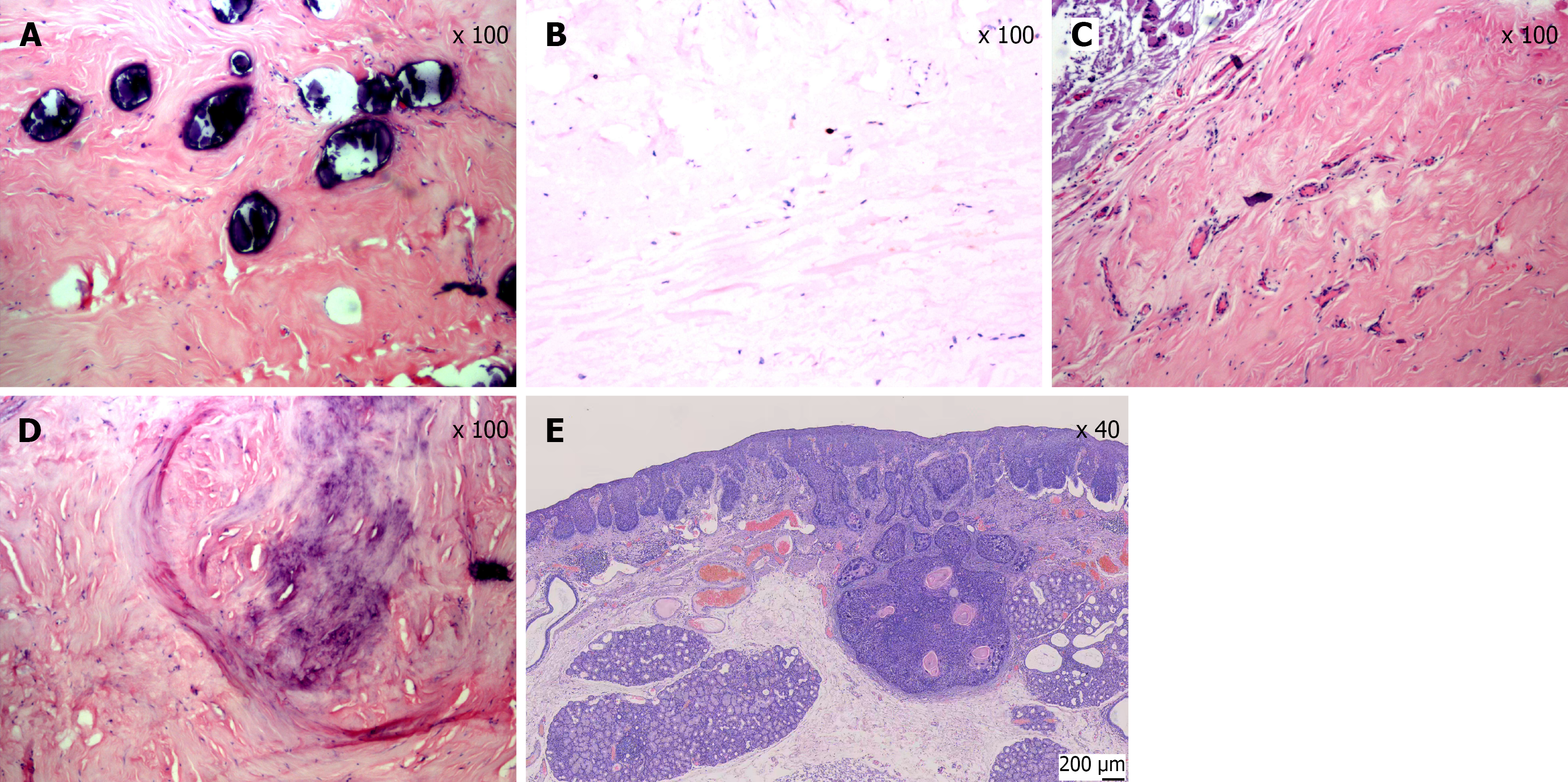
Figure 3 Histopathological and immunohistochemical features of the esophageal calcifying fibrous tumor and squamous cell carcinoma.
A: High-power hematoxylin and eosin (HE) staining (× 100) showing multiple basophilic calcified deposits within dense collagenous stroma. Scale bar = 100 μm; B: Immunohistochemical staining for Ki-67 (× 100) showing a low proliferation index (< 1%), consistent with a benign lesion. Scale bar = 100 μm; C: Low-power HE staining (× 100) showing paucicellular dense collagenous tissue with scattered bland spindle cells, without significant cytologic atypia or increased mitotic activity. Scale bar = 100 μm; D: High-power HE staining (× 100) showing chondroid-like hyalinized stroma with focal calcification. Scale bar = 100 μm; E: HE staining (× 40) of the mucosal lesion showing moderately differentiated esophageal squamous cell carcinoma invading the submucosa. Scale bar = 200 μm.
- Citation: Zhou Y, Duan H, Chen ZY, He DX. Endoscopic treatment of calcifying fibrous tumor of the esophagus combined with esophageal cancer: A case report. World J Gastrointest Oncol 2026; 18(11): 124574
- URL: https://www.wjgnet.com/1948-5204/full/v18/i11/124574.htm
- DOI: https://dx.doi.org/10.4251/wjgo.124574