
















Copyright: ©Author(s) 2026.
World J Gastrointest Oncol. Nov 15, 2026; 18(11): 124574
Published online Nov 15, 2026. doi: 10.4251/wjgo.124574
Published online Nov 15, 2026. doi: 10.4251/wjgo.124574
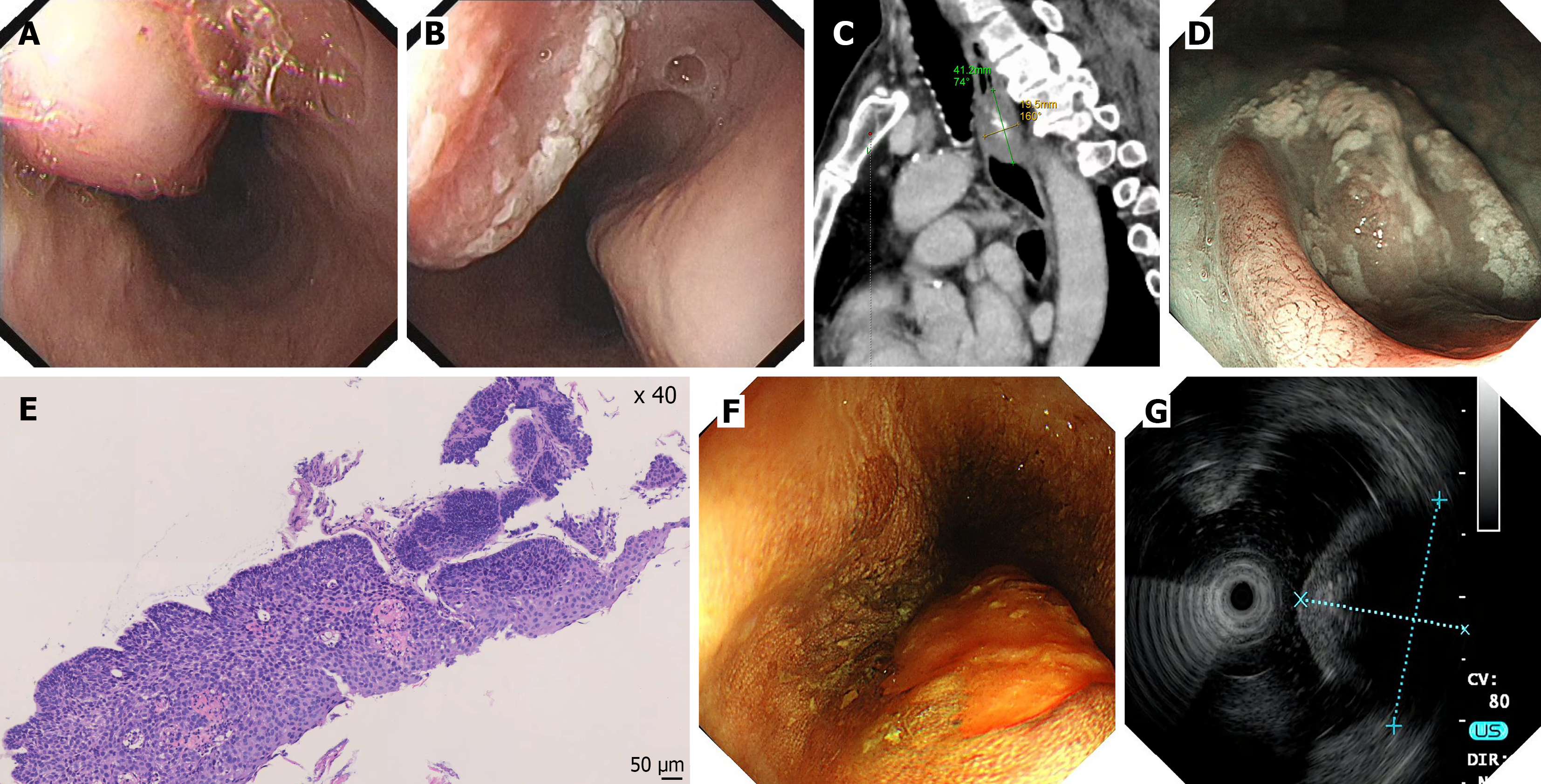
Figure 1 Preoperative multimodal evaluation of the esophageal lesions.
A: White-light endoscopy showing a submucosal tumor-like elevation in the upper esophagus; B: White-light endoscopy showing a superficially elevated mucosal lesion with grayish-white exudate on the surface; C: Contrast-enhanced chest computed tomography of the chest (sagittal view) shows calcification within the lesion; D: Magnifying narrow-band imaging endoscopy reveals type B2 intrapapillary capillary loops; E: Preoperative biopsy histopathology showing high-grade intraepithelial neoplasia of the esophageal squamous epithelium; F: Lugol iodine staining showing an unstained area of the lesion; G: Endoscopic ultrasonography shows the lesion as a hypoechoic mass with local hyperechoic areas.
- Citation: Zhou Y, Duan H, Chen ZY, He DX. Endoscopic treatment of calcifying fibrous tumor of the esophagus combined with esophageal cancer: A case report. World J Gastrointest Oncol 2026; 18(11): 124574
- URL: https://www.wjgnet.com/1948-5204/full/v18/i11/124574.htm
- DOI: https://dx.doi.org/10.4251/wjgo.124574