Copyright
©The Author(s) 2025.
World J Gastrointest Oncol. May 15, 2025; 17(5): 105448
Published online May 15, 2025. doi: 10.4251/wjgo.v17.i5.105448
Published online May 15, 2025. doi: 10.4251/wjgo.v17.i5.105448
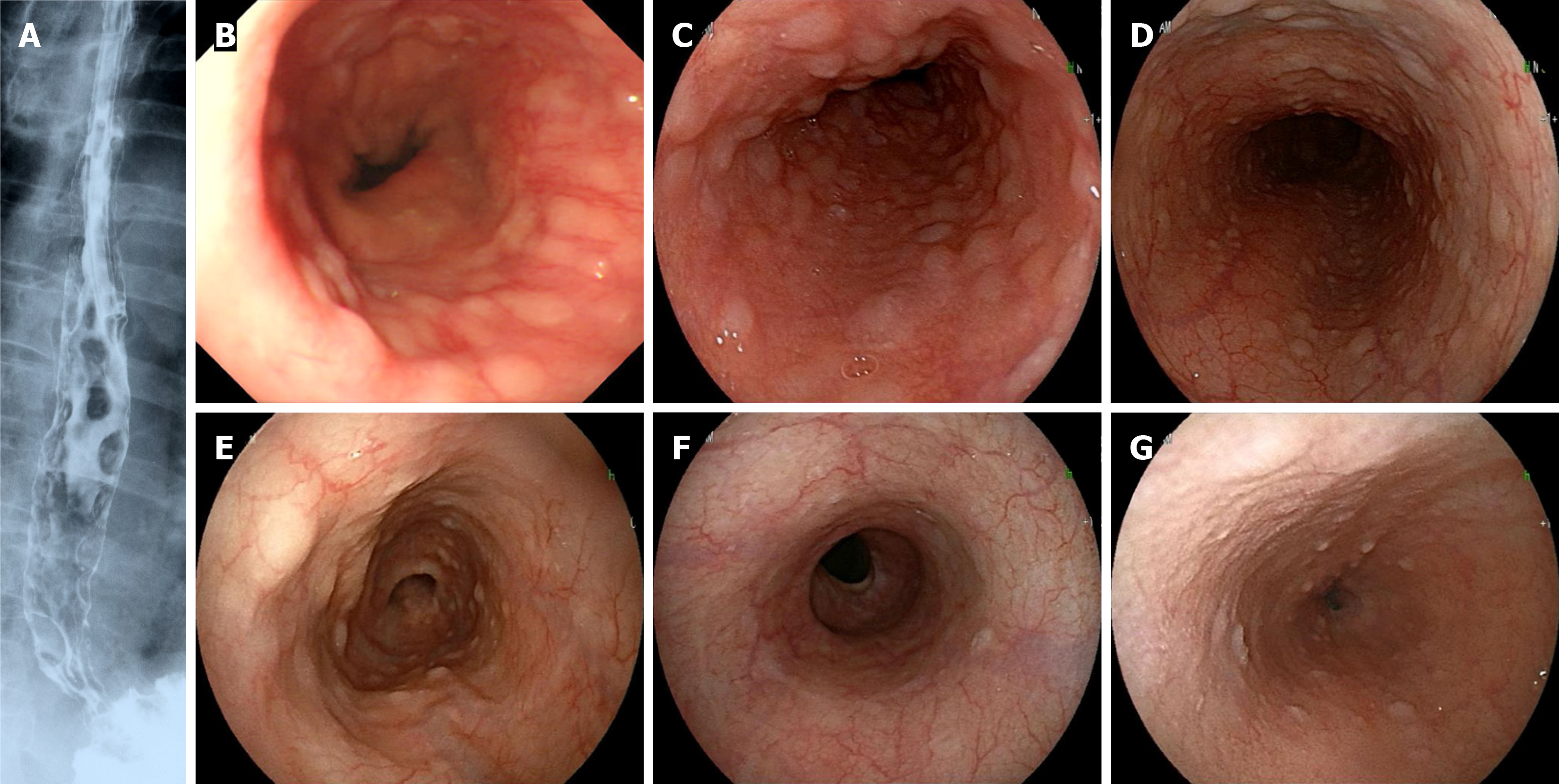
Figure 1 Esophageal findings of multiple lymphomatous polyposis.
A: Contrast X-rays (case 1); multiple lymphomatous polyposis was recognized from the middle to lower esophagus; B-D: Esophagogastroduodenoscopy of case 2 (B), case 3 (C) and case 4 (D); E-G: Before (E) and after (F) chemotherapy in case 5, and case 6 (G); In case 5, multiple lymphomatous polyposis (MLP) disappeared after treatment. In case 6, inconspicuous MLP findings were observed.
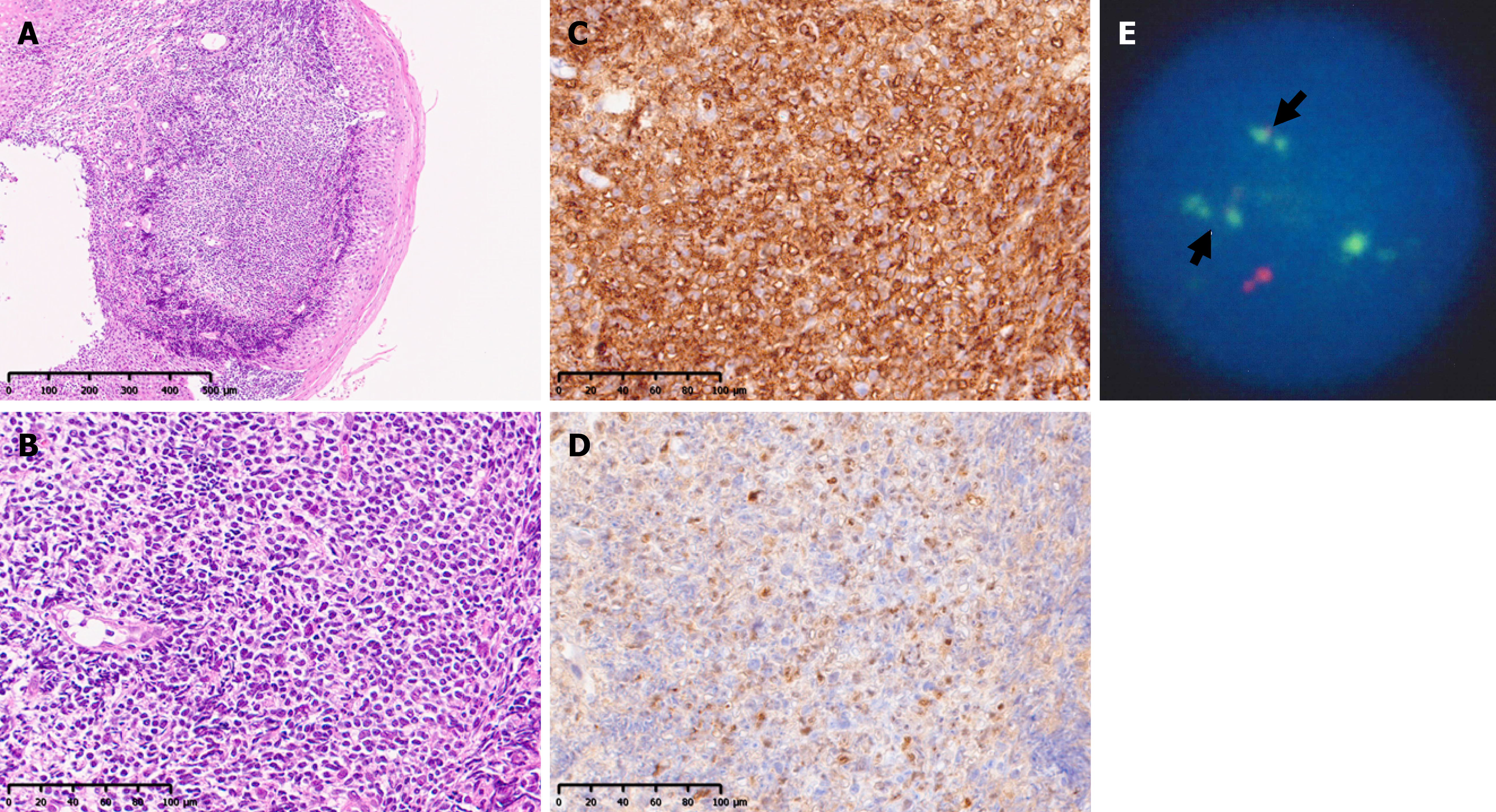
Figure 2 Histopathological findings and fluorescence in situ hybridization in case 6.
A: Low magnification (HE); B: High magnification (HE). Monotonous proliferation of small to medium-sized lymphoid cells was observed under the mucosa; C and D: CD5 (C) and cyclin D1 (D) was positive; E: IgH-BCL-1(CCND1) was strongly positive.
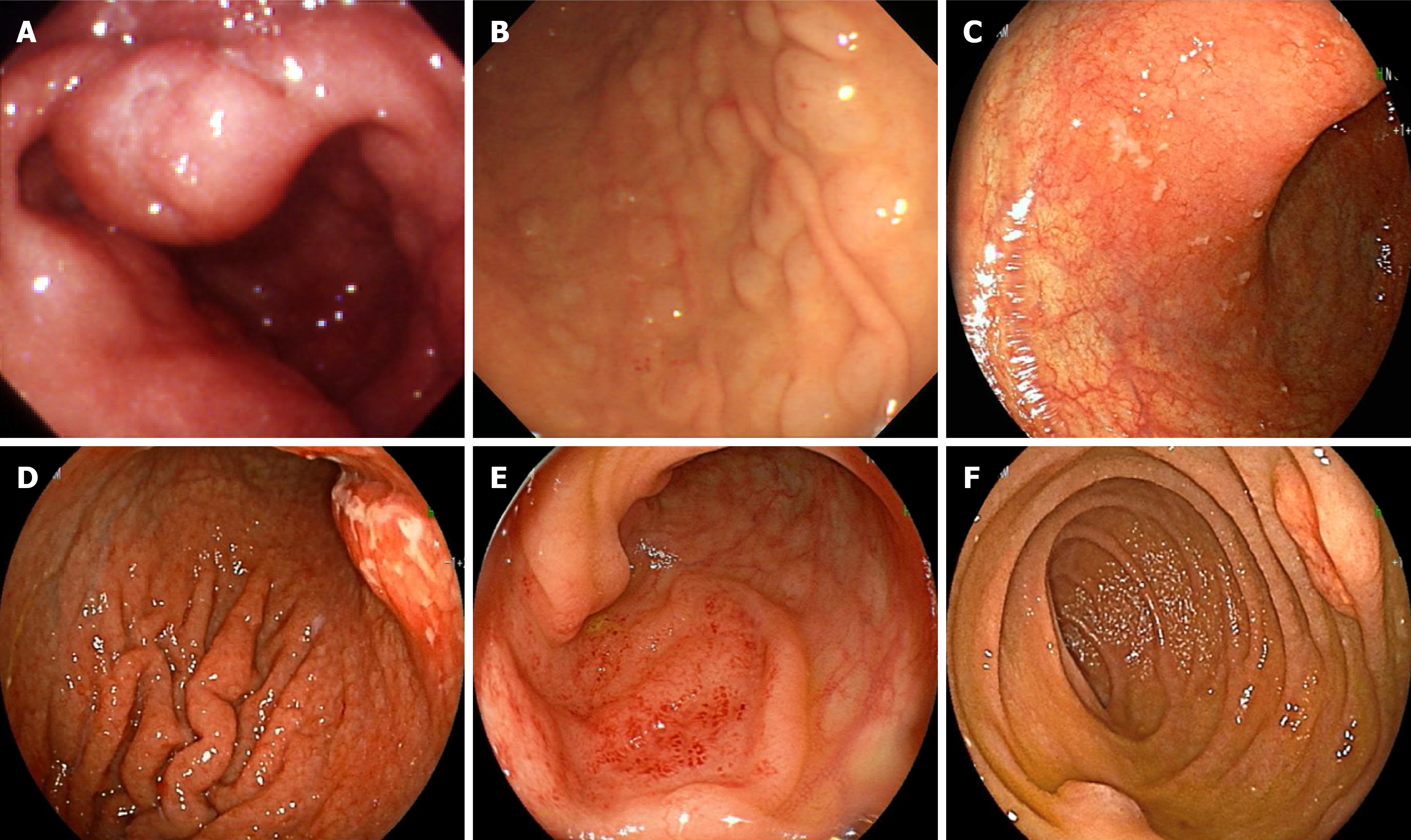
Figure 3 Endoscopic findings showing gastrointestinal involvement of mantle cell lymphoma.
A: Stomach in case 1, protruded type; B: Stomach in case 2, multiple lymphomatous polyposis (MLP) type; C: Stomach in case 3, superficial type; D: Stomach in case 4, fold thickening and protruded type; E: Ileum in case 5, protruded type; F: Duodenum in case 6, MLP type.
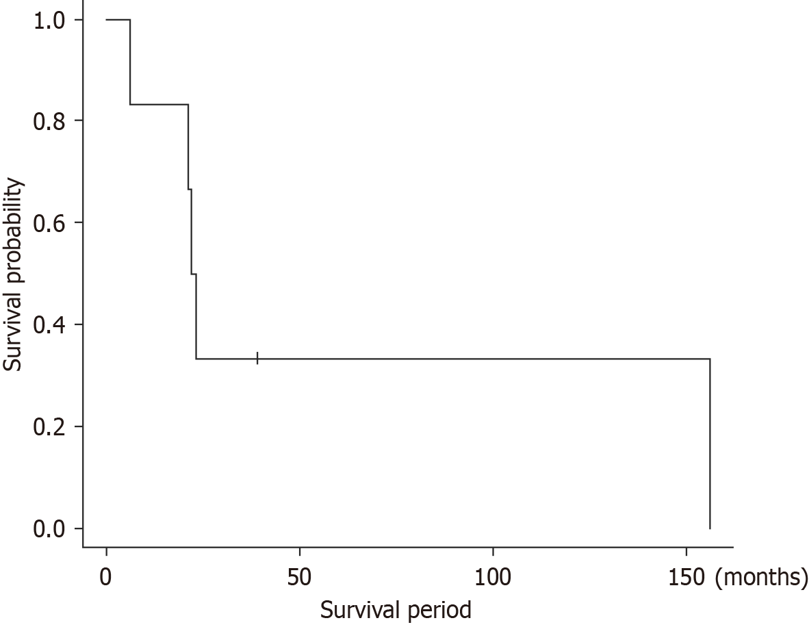
Figure 4 Overall survival curves for the 6 patients (Kaplan-Meier method).
The 50% survival period was less than 2 years, and the 5-year survival rate was approximately 30%.
- Citation: Saito M, Oda Y, Sugino H, Suzuki T, Yokoyama E, Kanaya M, Izumiyama K, Mori A, Morioka M, Kondo T. Esophageal involvement of mantle cell lymphoma presenting with multiple lymphomatous polyposis: A single-center study. World J Gastrointest Oncol 2025; 17(5): 105448
- URL: https://www.wjgnet.com/1948-5204/full/v17/i5/105448.htm
- DOI: https://dx.doi.org/10.4251/wjgo.v17.i5.105448


