Copyright
©The Author(s) 2025.
World J Gastrointest Oncol. May 15, 2025; 17(5): 105448
Published online May 15, 2025. doi: 10.4251/wjgo.v17.i5.105448
Published online May 15, 2025. doi: 10.4251/wjgo.v17.i5.105448
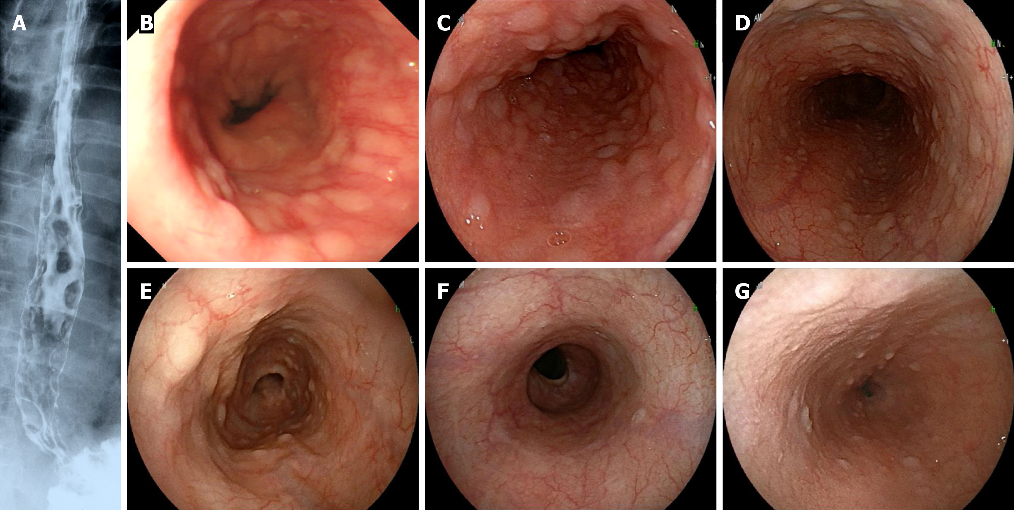
Figure 1 Esophageal findings of multiple lymphomatous polyposis.
A: Contrast X-rays (case 1); multiple lymphomatous polyposis was recognized from the middle to lower esophagus; B-D: Esophagogastroduodenoscopy of case 2 (B), case 3 (C) and case 4 (D); E-G: Before (E) and after (F) chemotherapy in case 5, and case 6 (G); In case 5, multiple lymphomatous polyposis (MLP) disappeared after treatment. In case 6, inconspicuous MLP findings were observed.
- Citation: Saito M, Oda Y, Sugino H, Suzuki T, Yokoyama E, Kanaya M, Izumiyama K, Mori A, Morioka M, Kondo T. Esophageal involvement of mantle cell lymphoma presenting with multiple lymphomatous polyposis: A single-center study. World J Gastrointest Oncol 2025; 17(5): 105448
- URL: https://www.wjgnet.com/1948-5204/full/v17/i5/105448.htm
- DOI: https://dx.doi.org/10.4251/wjgo.v17.i5.105448


