Copyright
©The Author(s) 2025.
World J Gastrointest Endosc. May 16, 2025; 17(5): 106074
Published online May 16, 2025. doi: 10.4253/wjge.v17.i5.106074
Published online May 16, 2025. doi: 10.4253/wjge.v17.i5.106074
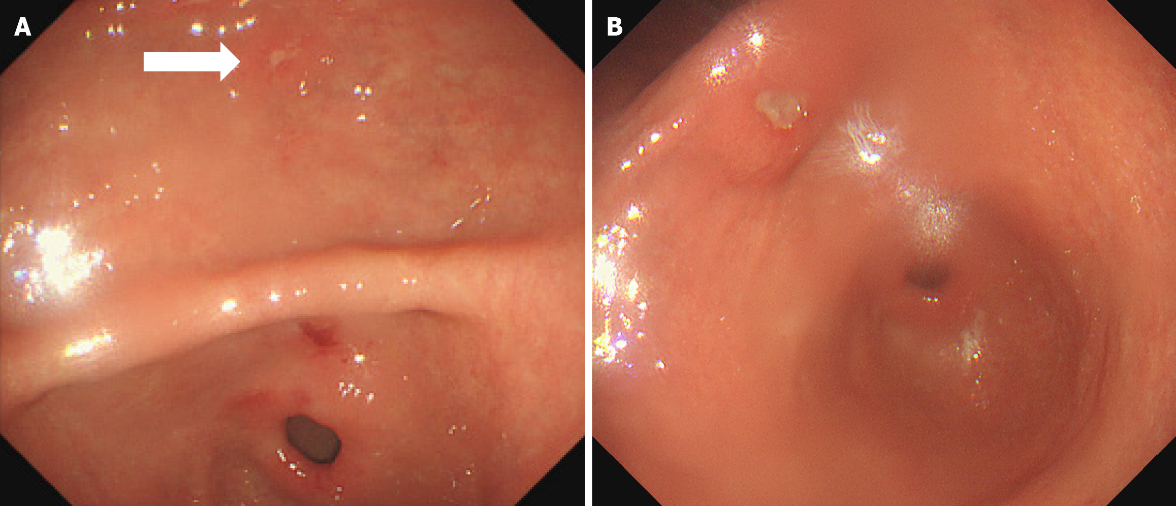
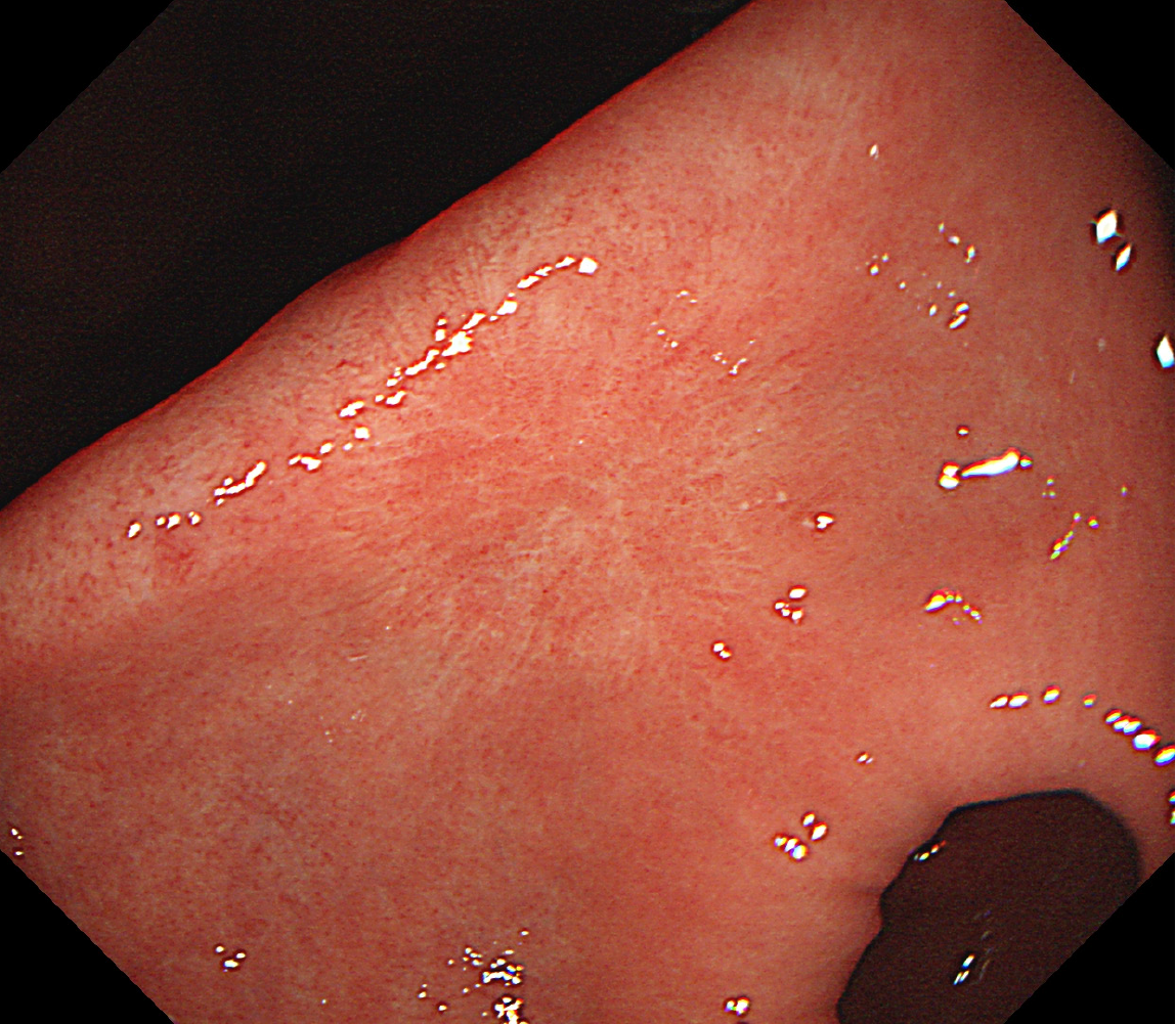
Figure 1 Endoscopic presentation of gastric antrum.
A: Multiple ulcers of the gastric antrum (white arrow) (2021-02-03); B: An ulcer on the small-curvature side of the antrum (2021-12-30).
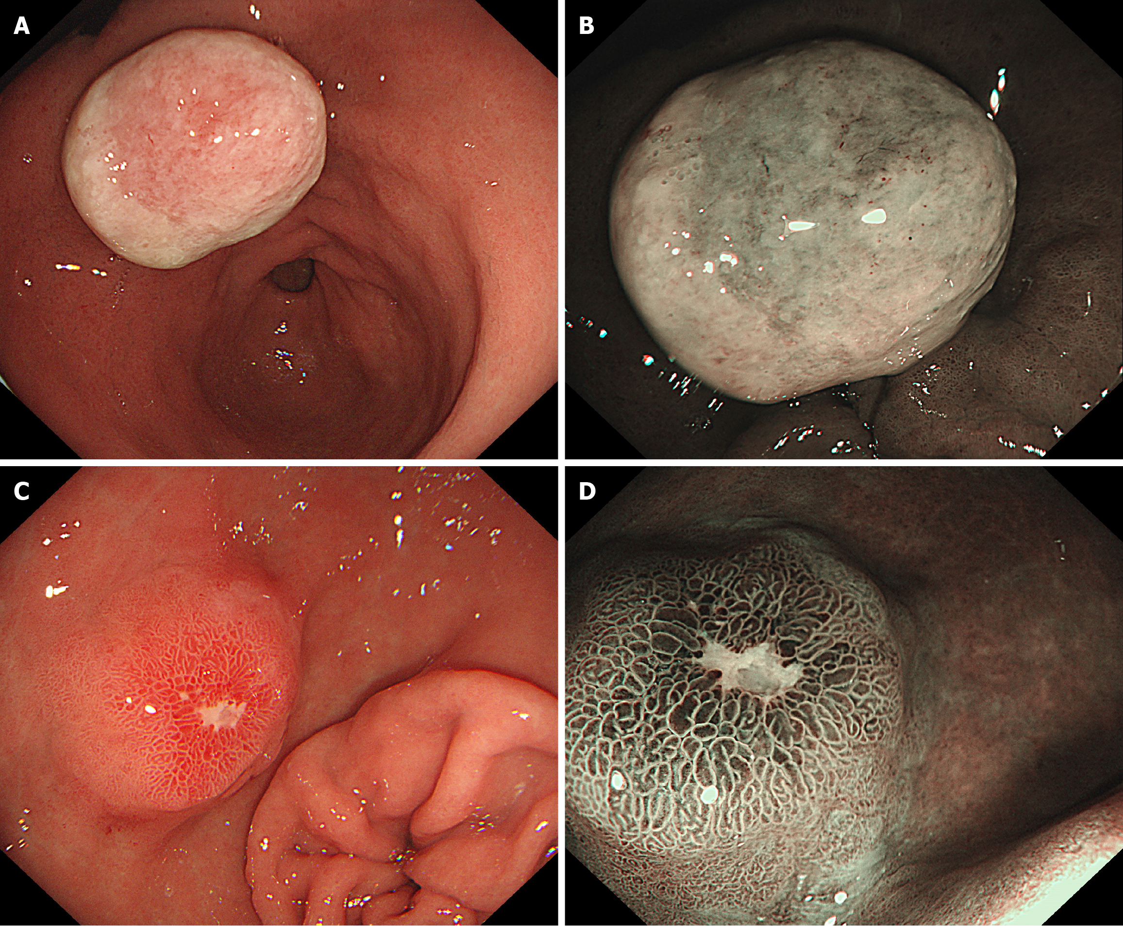
Figure 2 Comparison of proton pump inhibitors before and after treatment.
A: White light before proton pump inhibitors (PPIs) treatment: A 2 cm protrusion is seen in the lesser curvature of the gastric antrum, with surface coating attached; B: magnifying endoscopy with narrow-band imaging (ME-NBI) before PPIs treatment: Ulcer tissue attachment; C: White light after PPIs treatment: The bulge has shrunk compared to before, the surface of the lesion exhibited congestion, edema, erosion, hard texture upon palpation, and poor mobility; D: ME-NBI after PPIs treatment: Regular vascular pattern, normal pit pattern.
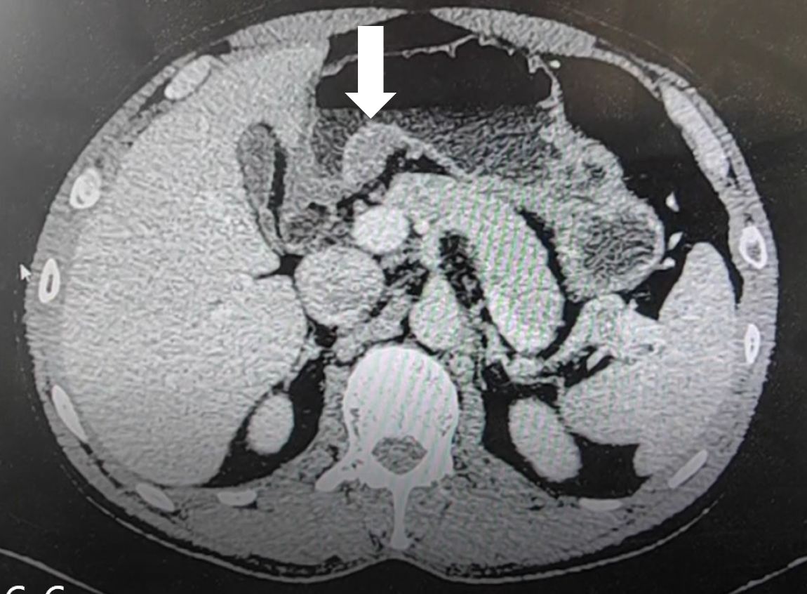
Figure 3 Gastric-enhanced computed tomography scan.
Gastric-enhanced computed tomography scan shows a soft tissue mass located beneath the mucosa on the small-curvature side of the antrum (white arrow).
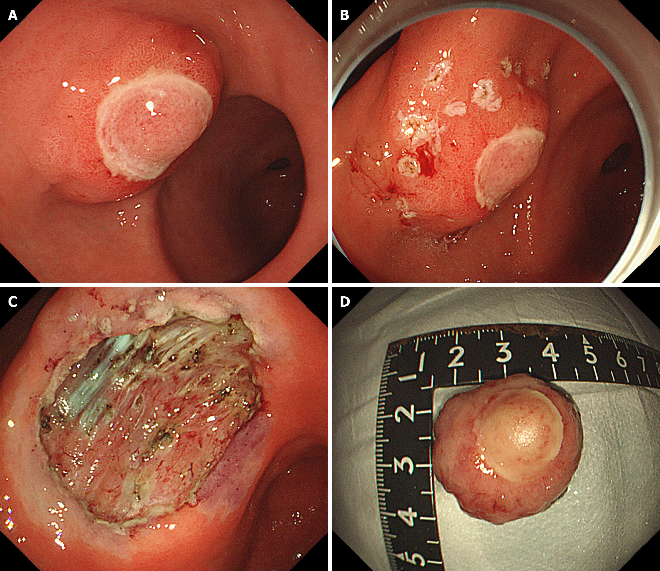
Figure 4 Endoscopic submucosal dissection.
A: The diameter of the submucosal bulge on the anterior wall of the gastric antrum was approximately 2.5 cm, with ulceration of the surface mucosa; B: Lateral mark of lesion; C: Postoperative wound; D: Postoperative specimen.
Figure 5 Gastroscopy examination after surgery.
A mucosal scar was seen in the gastric antrum 6 months after endoscopic treatment.
- Citation: Yang HC, Qu W. Diagnostic and therapeutic review of a rare gastric inflammatory fibroid polyps case with distinctive features: A case report. World J Gastrointest Endosc 2025; 17(5): 106074
- URL: https://www.wjgnet.com/1948-5190/full/v17/i5/106074.htm
- DOI: https://dx.doi.org/10.4253/wjge.v17.i5.106074


