Copyright
©The Author(s) 2025.
World J Gastrointest Endosc. May 16, 2025; 17(5): 106074
Published online May 16, 2025. doi: 10.4253/wjge.v17.i5.106074
Published online May 16, 2025. doi: 10.4253/wjge.v17.i5.106074
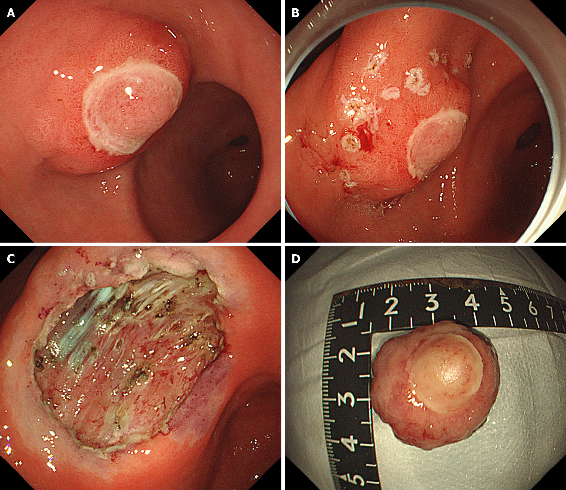
Figure 4 Endoscopic submucosal dissection.
A: The diameter of the submucosal bulge on the anterior wall of the gastric antrum was approximately 2.5 cm, with ulceration of the surface mucosa; B: Lateral mark of lesion; C: Postoperative wound; D: Postoperative specimen.
- Citation: Yang HC, Qu W. Diagnostic and therapeutic review of a rare gastric inflammatory fibroid polyps case with distinctive features: A case report. World J Gastrointest Endosc 2025; 17(5): 106074
- URL: https://www.wjgnet.com/1948-5190/full/v17/i5/106074.htm
- DOI: https://dx.doi.org/10.4253/wjge.v17.i5.106074


