Copyright
©The Author(s) 2025.
World J Hepatol. Jun 27, 2025; 17(6): 104073
Published online Jun 27, 2025. doi: 10.4254/wjh.v17.i6.104073
Published online Jun 27, 2025. doi: 10.4254/wjh.v17.i6.104073
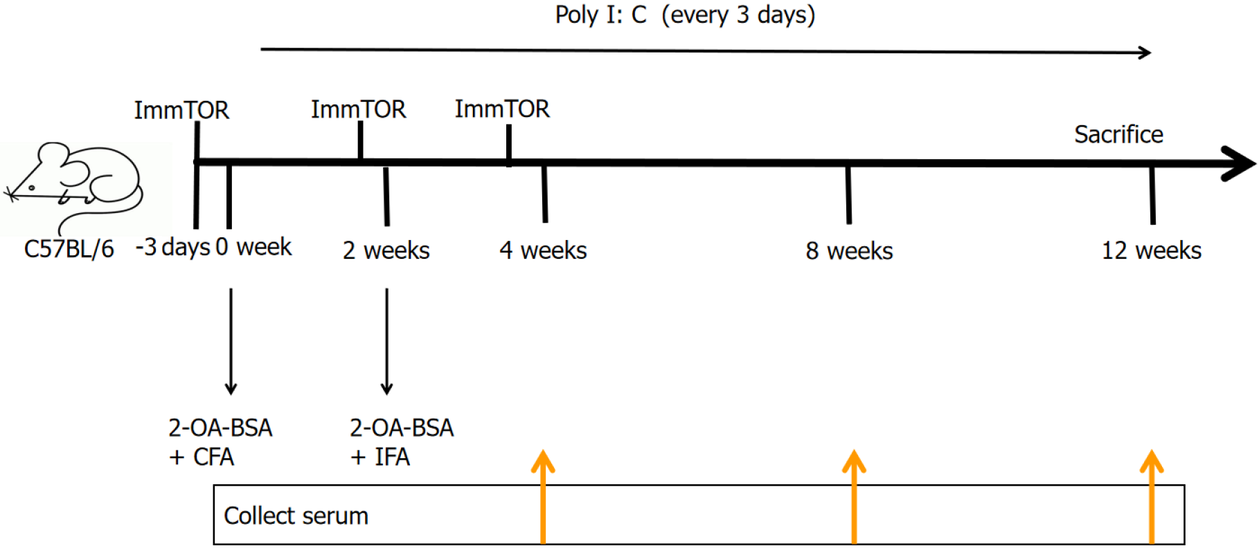
Figure 1 Animal modeling and intervention protocol.
CFA: Complete Freund's adjuvant; IFA: Incomplete Freund’s adjuvant; ImmTOR: Nanoparticles encapsulating rapamycin; Poly I: C: Polycytidylic acid; 2-OA-BSA: 2-octynoic acid-coupled bovine serum albumin.
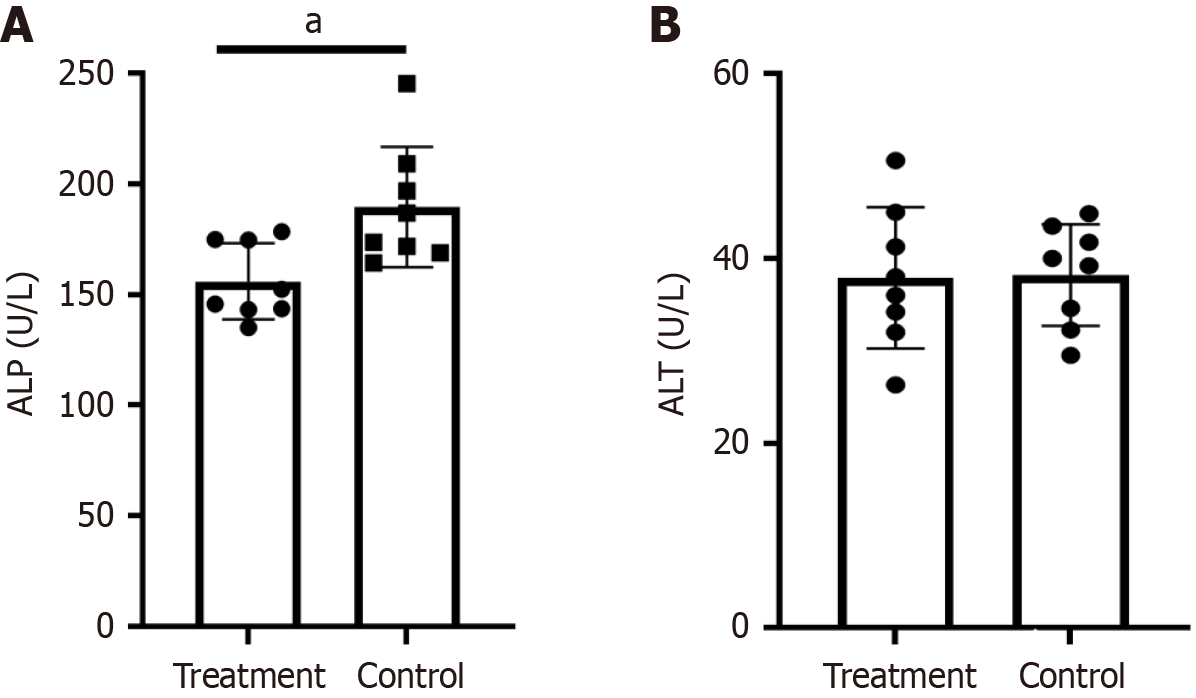
Figure 2 Analysis of biochemical parameters in serum in the treatment and control groups.
A: Nanoparticles encapsulating rapamycin treatment decreased alkaline phosphatase in the primary biliary cholangitis (PBC) mouse models (aP < 0.05); B: Comparison of alanine aminotransferase in the treatment and control group PBC mouse models. ALP: Alkaline phosphatase; ALT: Alanine aminotransferase.
Figure 3 Nanoparticles encapsulating rapamycin treatment decreased serum anti-mitochondrial antibodies levels and inhibited the expression of cytokines.
A: Levels of anti-mitochondrial antibodies at 4 weeks, 8 weeks and 12 weeks in primary biliary cholangitis (PBC) mice treated with nanoparticles encapsulating rapamycin and in mice in the control group; B: Comparative serum profiling of interferon gamma between control and treated PBC mice; C: Comparative serum levels of tumor necrosis factor α between the control and treatment group (aP < 0.05). AMA: Anti-mitochondrial antibodies; IFN-γ: Interferon gamma; TNF-α: Tumor necrosis factor α.
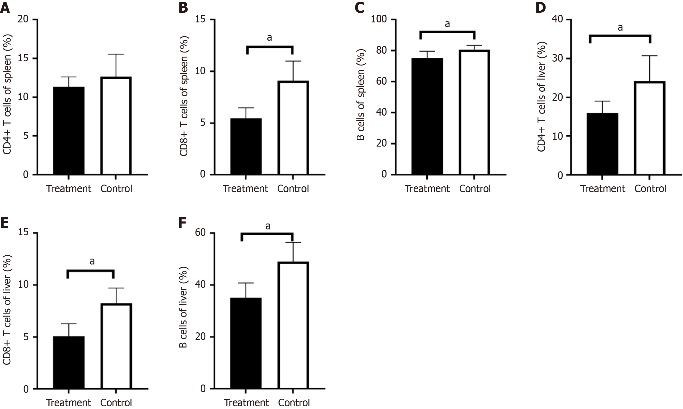
Figure 4 Nanoparticles encapsulating rapamycin treatment decreased the lymphocyte expression in the liver and spleen.
A: The percentage of CD4-positive T cells in the liver of the treatment group decreased compared with that in the control group; B: A comparison of the control and treatment groups showed that the percentage of CD8-positive T cells in the liver of the treatment group was decreased; C: The expression of B lymphocytes was decreased in the liver of the treatment group; D: There was no difference in CD4-positive T cells in the spleen between the two groups; E: The percentage of CD8-positive T lymphocytes in the spleen of the treatment group decreased compared with that in the control group; F: The expression of B lymphocytes in the spleen was decreased in the treatment group (aP < 0.05).
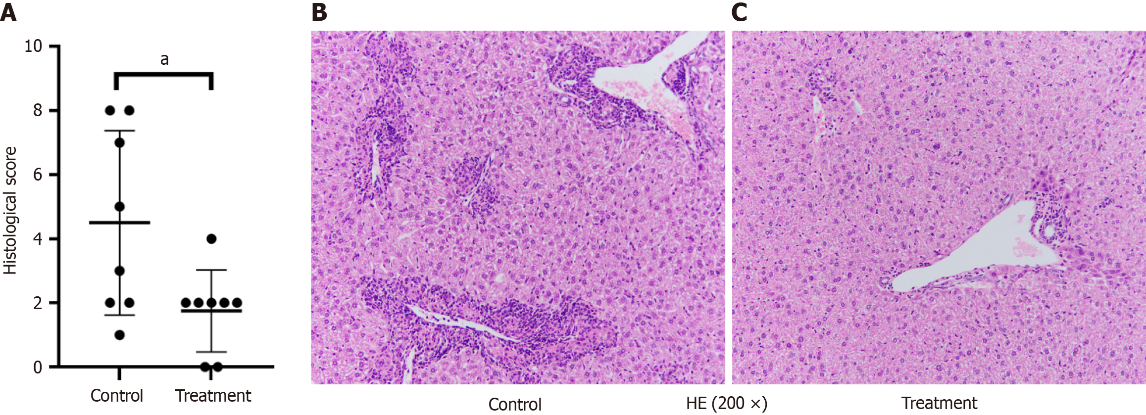
Figure 5 Nanoparticles encapsulating rapamycin treatment improved liver pathology.
A: Comparison of the hepatic histopathological score between the two groups (aP < 0.05); B: Hepatic histopathology visualized using hematoxylin and eosin staining in control mice; C: Hepatic pathological manifestations of mice in the treatment group. HE: Hematoxylin and eosin.
- Citation: Yang YS, Li XR, Wang ZM, Zheng L, Li JL, Cui XL, Song YB, Ma JJ, Guo HF, Gao LX, Zhou XH. Effect of rapamycin nanoparticles in an animal model of primary biliary cholangitis. World J Hepatol 2025; 17(6): 104073
- URL: https://www.wjgnet.com/1948-5182/full/v17/i6/104073.htm
- DOI: https://dx.doi.org/10.4254/wjh.v17.i6.104073


