Copyright
©The Author(s) 2025.
World J Stem Cells. Jun 26, 2025; 17(6): 107833
Published online Jun 26, 2025. doi: 10.4252/wjsc.v17.i6.107833
Published online Jun 26, 2025. doi: 10.4252/wjsc.v17.i6.107833
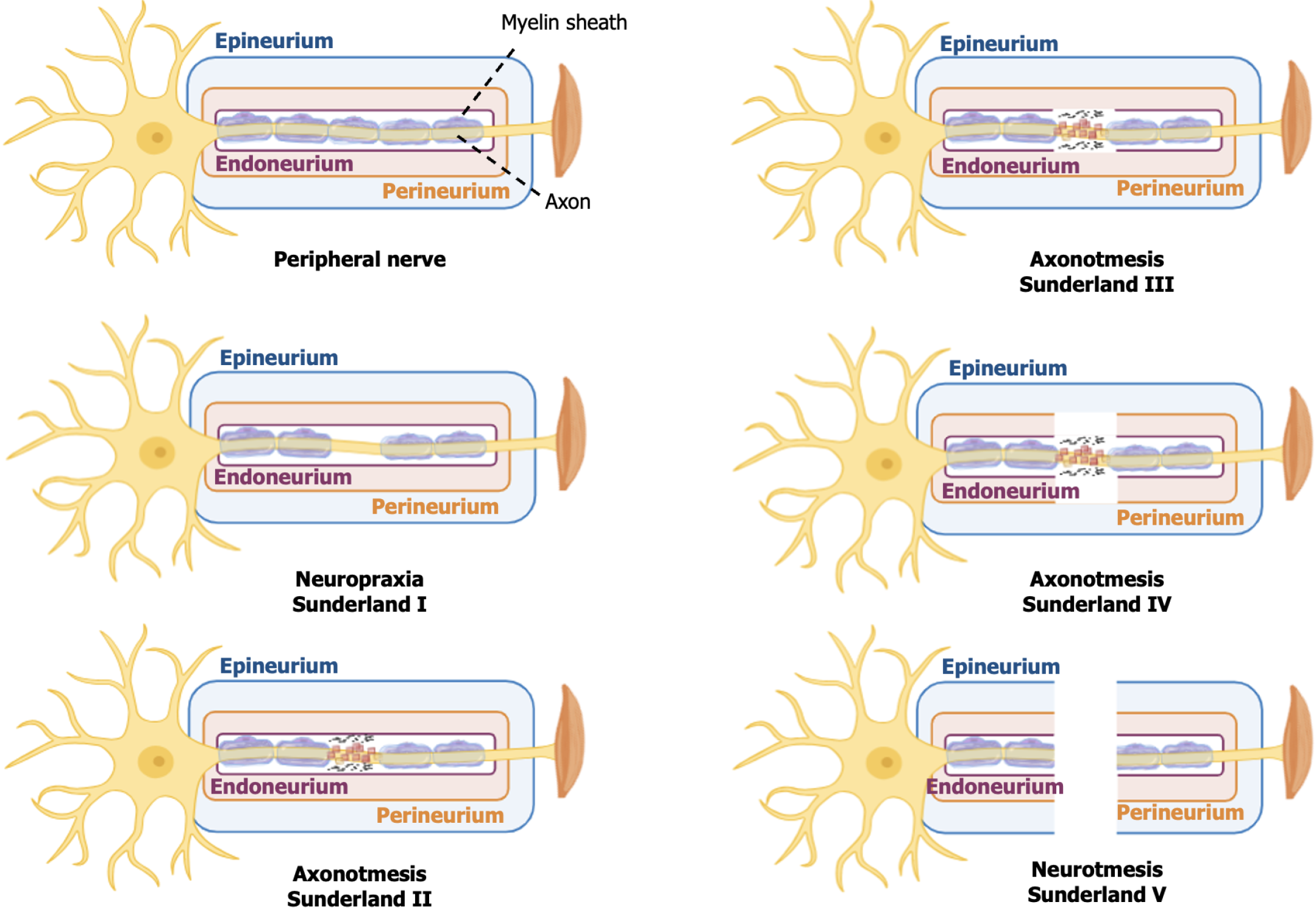
Figure 1 Schematic representation of the grading systems for peripheral nerve injuries.
Neuropraxia (Sunderland: Grade I) involves focal demyelination. Axonotmesis is subdivided into grade II (axon and myelin damage), grade III (axon, myelin, and endoneurium damage), and grade IV (axon, myelin, endoneurium, and perineurium damage). Neurotmesis (grade V) is complete nerve transection. Created in BioRender. Amorim, R. (2025) https://BioRender.com/z6uzrjj.
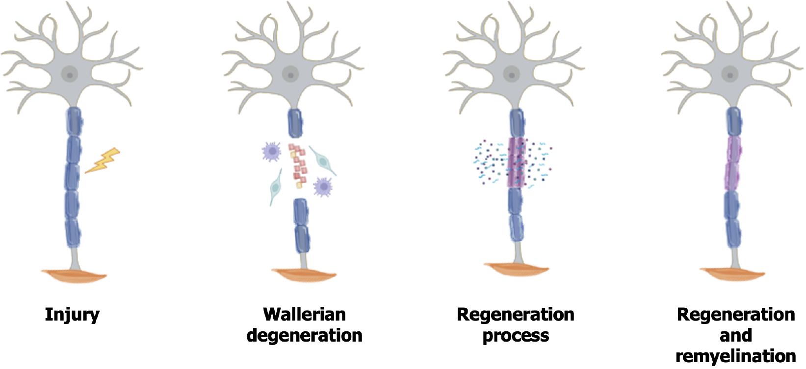
Figure 2 Endogenous regeneration process of the peripheral nervous system.
Following injury, Wallerian degeneration occurs, and Schwann cells proliferate and transdifferentiate while acting alongside macrophages to remove cellular and myelin debris. For regeneration Schwann cells form Büngner’s bands and secrete neurotrophic factors that support axonal growth. Created in BioRender. Amorim, R. (2025) https://BioRender.com/z6uzrjj.
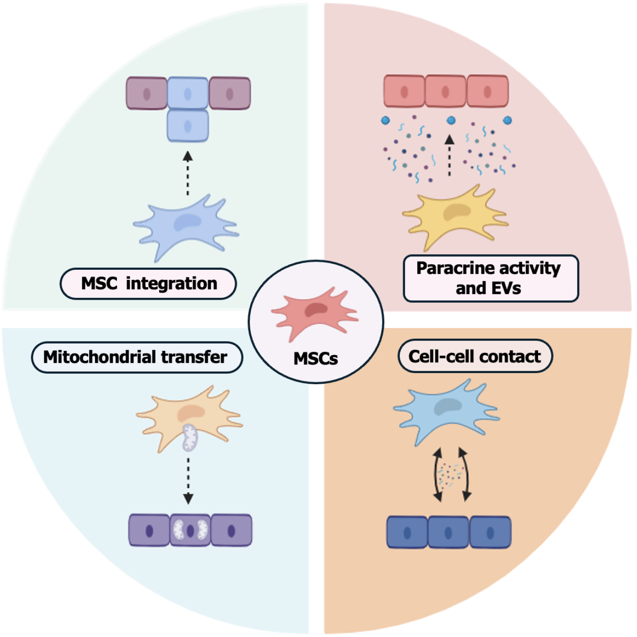
Figure 3 Schematic illustrating of the main mechanisms of mesenchymal stem cell action.
Mesenchymal stem cell integration, paracrine activity, release of extracellular vesicles, mitochondrial transfer, and cell-cell contact. Created in BioRender. Amorim, R. (2025) https://BioRender.com/z6uzrjj.MSCs: Mesenchymal stem cells; EVs: Extracellular vesicles.
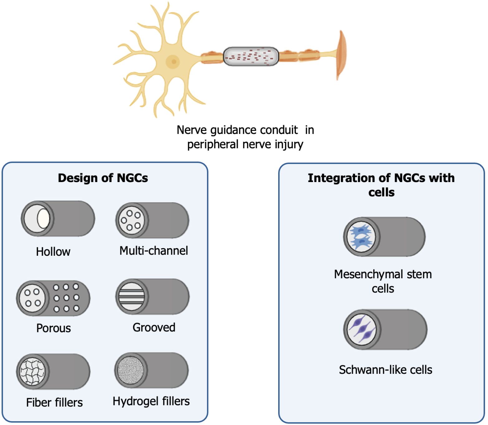
Figure 4 Summarized schematic of different designs to enhance nerve guidance conduits (NGCs).
Design variations include hollow tubes, multichannel, porous, grooved structures, fiber, or hydrogel fillers. These designs can be used alone or in combination with mesenchymal stem cells or Schwann-like cells for the treatment of peripheral nerve injuries. NGCs: Nerve guidance conduits. Created in BioRender. Amorim, R. (2025) https://BioRender.com/z6uzrjj.
- Citation: Ferreira LVO, Roballo KCS, Amorim RM. Mesenchymal stem cell-based therapy for peripheral nerve injuries: A promise or reality? World J Stem Cells 2025; 17(6): 107833
- URL: https://www.wjgnet.com/1948-0210/full/v17/i6/107833.htm
- DOI: https://dx.doi.org/10.4252/wjsc.v17.i6.107833


