Published online Feb 14, 2018. doi: 10.3748/wjg.v24.i6.744
Peer-review started: December 8, 2017
First decision: December 21, 2017
Revised: January 3, 2018
Accepted: January 16, 2018
Article in press: January 16, 2018
Published online: February 14, 2018
Processing time: 59 Days and 21.2 Hours
To describe contrast-enhanced ultrasound (CEUS) features and evaluate differential diagnosis value of CEUS and conventional ultrasound for patients with benign and malignant gallbladder lesions.
This study included 105 gallbladder lesions. Before surgical resection and pathological examination, conventional ultrasound and CEUS were performed to examine for lesions. Then, all the lesions were diagnosed as (1) benign, (2) probably benign, (3) probably malignant or (4) malignant using both conventional ultrasound and CEUS. The CEUS features of these gallbladder lesions were analyzed and diagnostic efficiency between conventional ultrasound and CEUS was compared.
There were total 17 cases of gallbladder cancer and 88 cases of benign lesion. Some gallbladder lesions had typical characteristics on CEUS (e.g., gallbladder adenomyomatosis had typical characteristics of small nonenhanced areas on CEUS). The sensitivity, specificity, positive predictive value, negative predictive value and accuracy of CEUS were 94.1%, 95.5%, 80.0%, 98.8% and 95.2%, respectively. These were significantly higher than conventional ultrasound (82.4%, 89.8%, 60.9%, 96.3% and 88.6%, respectively). CEUS had an accuracy of 100% for gallbladder sludge and CEUS helped in differential diagnosis among gallbladder polyps, gallbladder adenoma and gallbladder cancer.
CEUS may provide more useful information and improve the diagnosis efficiency for the diagnosis of gallbladder lesions than conventional ultrasound.
Core tip: With the advent of ultrasound contrast agents, contrast-enhanced ultrasound (CEUS) is playing a more and more important role clinically. However, the value of CEUS in gallbladder lesions has not been widely accepted yet. In this study, we evaluated the differential diagnosis value of CEUS and conventional ultrasound for patients with benign and malignant gallbladder lesions. Our results showed that CEUS may provide more useful information and improve diagnosis efficiency for the diagnosis of gallbladder lesions than conventional ultrasound.
- Citation: Zhang HP, Bai M, Gu JY, He YQ, Qiao XH, Du LF. Value of contrast-enhanced ultrasound in the differential diagnosis of gallbladder lesion. World J Gastroenterol 2018; 24(6): 744-751
- URL: https://www.wjgnet.com/1007-9327/full/v24/i6/744.htm
- DOI: https://dx.doi.org/10.3748/wjg.v24.i6.744
Conventional ultrasound is the primary and most important imaging modality for gallbladder diseases. The excellent image contrast between anechoic bile and gallbladder wall or gallbladder diseases, and the increasingly improved ultrasound spatial resolution ensure conventional ultrasound to have a high detection rate of gallbladder diseases[1]. With the advantages of real-time imaging, safety with no radiation, great cost effectiveness and great spatial resolution, conventional ultrasound makes itself more suitable than computed tomography (CT) and magnetic resonance imaging (MRI) for the detection of gallbladder diseases[2].
Despite the above-mentioned advantages of conventional ultrasound, the sensitivity and accuracy are not satisfactory, especially when stones or some other gallbladder lesions fill the gallbladder lumen[3,4]. With no information of microvascularity, it is very hard to differentiate some benign diseases, such as benign gallbladder wall thickening or motionless sludge, from malignant ones using conventional ultrasound. The application of microbubbles could help in the differential diagnosis by providing useful perfusion information in the lesions[5].
Contrast-enhanced ultrasound (CEUS) has been widely used in liver disease, with an excellent diagnostic efficiency comparable to contrast-enhanced CT[6-8]. The value of CEUS in other organs, such as kidney, breast, etc., has also been well established and identified[9,10]. Although the value in gallbladder has not been recognized and accepted by the European Federation of Societies for Ultrasound in Medicine and Biology[11], there have been some studies which have shown the usefulness of CEUS in the differential diagnosis between benign and malignant gallbladder lesions[5,12].
In this study, we described CEUS features and evaluated differential diagnosis value of CEUS and conventional ultrasound for patients with benign and malignant gallbladder lesions.
The Ethics Committee of our hospital approved this study. Before the sonographic examination, we obtained all patients’ written informed consent. The features of gallbladder lesions in CEUS were analyzed and described retrospectively. The study and comparison of the diagnostic efficiency between CEUS and conventional ultrasound was designed prospectively.
Between December 2012 and October 2016, 136 gallbladder lesions in 133 patients were imaged using both conventional ultrasound and CEUS in our hospital. Of these, 31 lesions were excluded from this study because the patients did not undergo cholecystectomy and were without pathological diagnosis. Therefore, 105 gallbladder lesions in 103 patients (47 males and 56 females; mean age ± standard deviation, 42.5 ± 10.6 years) were included in this study.
All the conventional ultrasound and CEUS examinations were performed by an ultrasound physician with thirteen years’ experience in conventional ultrasound and five years’ experience in CEUS. An Acuson S2000 diagnostic ultrasound system or an Acuson Sequoia 512 diagnostic ultrasound system (Siemens Medical Solutions, Mountain View, CA, United States) equipped with a transabdominal curvilinear transducer running on CadenceTM Contrast pulse sequence (CPS) software were used for all the ultrasound examinations. All the patients fasted at least for 8 h before the examinations.
Conventional ultrasound examinations were first performed to detect the gallbladder lesions. The lesion’s size, location, shape, stalk, boundary, echogenicity and wall destruction were analyzed and recorded. Then, Doppler vascularity was observed using color Doppler ultrasound. A diagnosis of benign, probably benign, probably malignant or malignant was made according to conventional ultrasound features, by two radiologists with at least ten years’ experience in both conventional ultrasound and CEUS. If they concluded different diagnosis, a third radiologist (with twenty-five years’ experience in conventional ultrasound and twelve years’ experience in CEUS) discussed together with them and decided on a final diagnosis.
For CEUS examinations, the same ultrasound machines were used. SonoVue (Bracco, Italy), the only microbubbles permitted for clinical use in China, was used in this study and was prepared following the appropriate guidelines before examinations. Every patient was instructed to take gentle and steady breaths to minimize the influence by respiratory movement. When the target lesion was shown clearly using conventional ultrasound, the CPS mode (MI: 0.21) was activated. A dose of 1.6 mL of SonoVue was administrated through the antecubital vein as a bolus immediately followed by 5 mL 0.9% saline solution. A stopwatch was started at the same time. The image was observed and recorded for 2 min and then the whole gallbladder and the liver were scanned to find other lesions and liver infiltration. After that, CEUS features of the lesion were analyzed and a diagnosis of benign, probably benign, probably malignant or malignant was made according to CEUS features by the above-mentioned radiologists.
After the resection of gallbladder lesions and the final pathological diagnosis was made, CEUS images were reviewed and the features of each kind of gallbladder lesions in CEUS were analyzed and summarized.
SPSS version 13.0 software (IBM Corporation, Chicago, IL, United States) was used for statistical analysis. P < 0.05 was considered a statistically significant difference. The diagnostic efficiency of conventional ultrasound and CEUS was assessed in terms of sensitivity, specificity, positive predictive value, negative predictive value and accuracy and was compared using chi-square test and Fisher’s exact test.
There were 17 malignant and 88 benign gallbladder lesions in total in this study according to the histopathological diagnosis after cholecystectomy, including 17 cases of gallbladder cancer, 11 case of gallbladder sludge, 28 cases of gallbladder adenomyomatosis, 36 cases of gallbladder polyps and 13 cases of gallbladder adenoma.
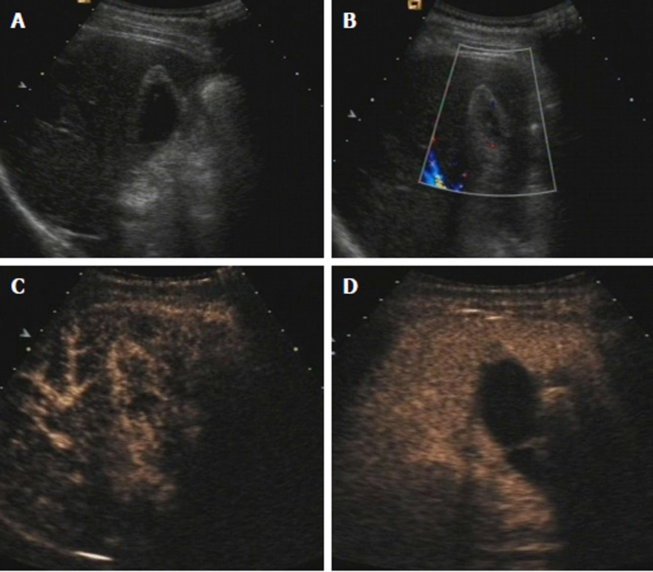
All the cases of gallbladder sludge were shown as completely nonenhanced on CEUS, and the diagnostic accuracy was 100% (Figure 1).
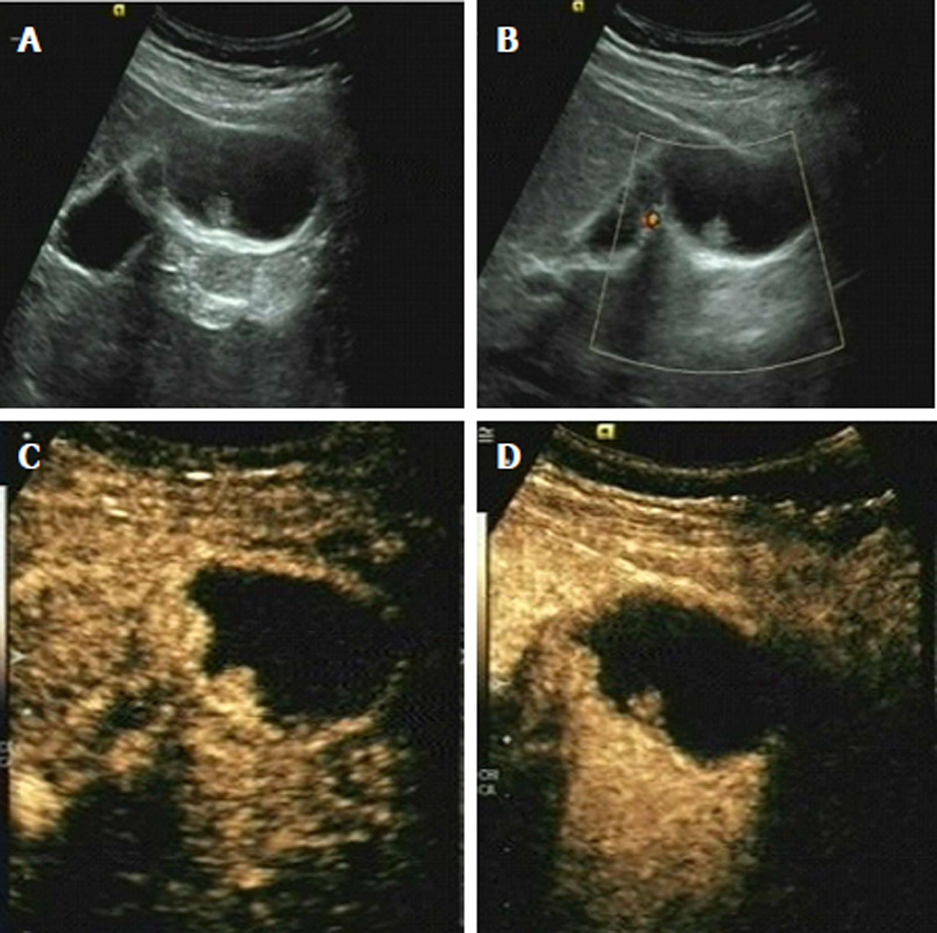
Gallbladder adenomyomatosis was mostly shown as heterogeneously enhanced, with some small nonenhanced areas (represented as Rokitansky-Aschoff sinuses) on both arterial phase and venous phase (Figure 2). Some of them were together with echogenic foci and tail sign.
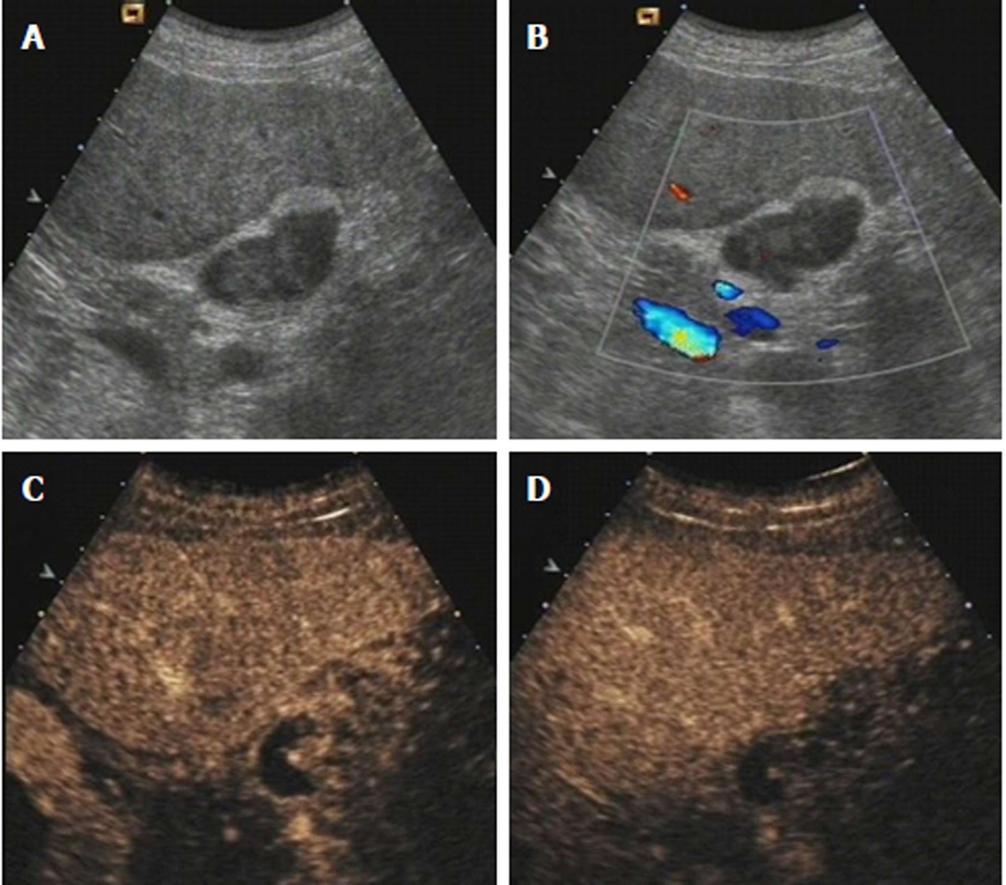
Gallbladder polyps and gallbladder adenoma were mostly shown as homogeneously hyperenhanced on arterial phase and isoenhanced on venous phase. The gallbladder wall was intact and the surrounding tissue was normal, with no invasion (Figure 3).
The appearances of gallbladder cancer on CEUS were various. It could be a mass in gallbladder which was heterogeneously hyperenhanced on arterial phase and washed out quickly (Figure 4). Or, the irregular thickness of gallbladder, which was also heterogeneously hyperenhanced on arterial phase and washed out quickly, could be a sign of malignancy. In some cases, the intact gallbladder wall was destroyed or the surrounding liver tissue was invaded.
Besides providing microvascular information, CEUS makes the contour of a lesion much clearer and the evaluation of a lesion’s shape, size and boundary much more accurate.
The diagnostic results of conventional ultrasound are shown in Table 1. There were 3 malignant lesions misdiagnosed as probably benign and 5 diagnosed as probably malignant. There were 8 benign lesions (2 cases of sludge, 3 cases of adenomyomatosis, 2 cases of polyps and 1 case of gallbladder adenoma) misdiagnosed as probably malignant, and one benign lesion misdiagnosed as definitely malignant (1 case of adenoma). A total of 18 benign lesions (3 cases of sludge, 5 cases of adenomyomatosis, 5 cases of polyps and 5 cases of gallbladder adenoma) were diagnosed as probably benign.
| Benign | Malignant | |||
| definitely | probably | probably | definitely | |
| Benign, n = 88 | 61 (69.3) | 18 (20.5) | 8 (9.1) | 1 (1.1) |
| Malignant, n = 17 | 0 (0) | 3 (17.6) | 5 (29.4) | 9 (52.9) |
The sensitivity, specificity, positive predictive value, negative predictive value and accuracy of conventional ultrasound were shown in Table 2.
| Features of lesions | Sensitivity | Specificity | Positive predictive value | Negative predictive value | Accuracy |
| Conventional ultrasound | 82.4 | 89.8 | 60.9 | 96.3 | 88.6 |
| Contrast-enhanced ultrasound | 94.1 | 95.5 | 80.0 | 98.8 | 95.2 |
| P value | 0.301 | 0.124 | 0.152 | 0.297 | 0.064 |
The diagnostic results of CEUS are shown in Table 3. Two malignant lesions which were misdiagnosed as probably benign by conventional ultrasound were correctly diagnosed as probably malignant by CEUS, and one malignant lesion which was diagnosed as probably malignant by conventional ultrasound was confirmed as malignant by CEUS. For benign lesions, all the cases of sludge were confirmed as benign. All cases of adenomyomatosis but 3 (1 diagnosed as probably malignant and 2 as probably benign) and all cases of polyps but 3 (1 diagnosed as probably malignant and 2 as probably benign) were confirmed as benign. Two cases of adenoma were misdiagnosed as probably malignant and another two cases of adenoma were diagnosed as probably benign. The rest 9 of the cases of adenoma were confirmed as benign.
| Benign | Malignant | |||
| Definitely | Probably | Probably | Definitely | |
| Benign, n = 88 | 78 (88.6) | 6 (6.8) | 4 (4.5) | 0 (0) |
| Malignant, n = 17 | 0 (0) | 1 (5.9) | 6 (35.3) | 10 (58.8) |
The sensitivity, specificity, positive predictive value, negative predictive value and accuracy of CEUS are shown in Table 2. The diagnostic efficiencies of CEUS were all significantly higher than those of conventional ultrasound, though the differences were not statistically significant.
In this study, we compared the value of CEUS in the differential diagnosis of benign and malignant gallbladder lesions with conventional ultrasound. Our results showed that the diagnostic efficiencies of CEUS were much higher than those of conventional ultrasound, though the differences were not statistically significant. With all the advantages and information of conventional ultrasound, CEUS provides more information about the important microvascularity in lesions. Also, with the application of microbubbles, the contour, the boundary and the shape of a lesion, the intactness of gallbladder wall and the invasion of the surrounding tissue could be revealed more clearly. So, the diagnostic efficiencies were highly improved, though the differences between the diagnostic efficiencies were not statistically significant.
Although the clinical significance of gallbladder sludge has not been confirmed yet, the accurate diagnosis is still of importance to avoid unnecessary examination and treatment[13]. Gallbladder sludge is usually shown on ultrasound as movable, echogenic matter, which could be easily diagnosed. However, sometimes gallbladder sludge could be shown as an intraluminal mass and imitates tumors such as gallbladder cancer or adenoma[14]. Then, the differential diagnosis is very difficult using conventional ultrasound. CEUS is very useful at such a time. As sludge has no blood supply inside it, it shows a complete nonenhancement on both arterial phase and venous phase. The diagnostic accuracy was 100% in our study, and the result was similar with some previous studies[11,15].
Gallbladder adenomyomatosis is a noninfectious and nontumorous disease of gallbladder which is usually found accidentally, with no malignant potential and which needs no specific treatment[16]. It has some typical characteristics on CEUS, too. With the small nonenhanced areas on arterial phase and venous phase (represented as Rokitansky-Aschoff sinuses), together with echogenic foci and tail sign or not, the correct diagnosis would be easily made[17,18]. The study by Tang et al[17] showed that small anechoic spaces or intramural echogenic foci were 100% detected using CEUS, which made the diagnostic accuracy much higher than conventional ultrasound. In this study, besides one case with no small anechoic spaces that was misdiagnosed as probably malignant, the rest of the cases were all diagnosed correctly as gallbladder adenomyomatosis.
The differential diagnosis among gallbladder polyps, gallbladder adenoma and gallbladder cancer was not easy on CEUS. However, some studies showed that some CEUS features were useful and significant for differentiating malignancy from benignity. The study of Xu et al[19] showed that focal gallbladder wall thickening, inner layer discontinuity and outer layer discontinuity were associated with gallbladder malignancy. Branched or linear intralesional vessels, tortuous-type tumor vessel, enhanced heterogeneously in the artery phase and washed out quickly in the late phase were usually considered as signs for malignancy[20-22]. On the contrary, gallbladder polyps or gallbladder adenoma was usually enhanced homogenously and the microbubbles inside the lesions washed out together with normal gallbladder wall. Recently, the study of the differential diagnosis of localized gallbladder lesions using contrast-enhanced harmonic endoscopic ultrasonography also confirmed the value of CEUS for the evaluation and differentiation of localized gallbladder lesions[23]. Although CEUS provides the microvascular information, conventional ultrasound is still very important and is the foundation of CEUS. The size, shape and boundary of a lesion, the intactness of gallbladder wall and the invasion of surrounding tissue are very important for the differential diagnosis. Besides providing microvascular information, CEUS makes the contour of a lesion much clearer and the evaluation of a lesion’s shape, size and boundary much more accurate. That is an important reason for the improvement of CEUS diagnostic efficiency, compared with conventional ultrasound.
Our study has some limitations. First, the sample was not large enough, especially for the malignant lesions. The pathological types of the lesions were not enough in number, either. For example, all the gallbladder adenomyomatosis in our study were of localized type, and no segmental or diffuse types were included. And, there were only a few early-stage cancers in this study, making it hard to compare the difference between benign lesions and early-stage cancers on CEUS. Second, the CEUS features were not analyzed using quantitative analysis software, but by naked eyes. No quantitative parameters were acquired and analyzed. Furthermore, the interobserver agreement in CEUS and conventional ultrasound was not compared in this study.
In conclusion, gallbladder sludge and gallbladder adenomyomatosis had special features on CEUS and the diagnostic accuracy was very high. CEUS helped the differential diagnosis among gallbladder polyps, gallbladder adenoma and gallbladder cancer. The diagnostic efficiency of CEUS was highly improved compared to conventional ultrasound.
With the advent of ultrasound contrast agents, contrast-enhanced ultrasound (CEUS) is playing a more and more important role clinically. CEUS is a safe, convenient and repeatable imaging method, with no risk of serious allergy and radiation. CEUS has an excellent diagnostic efficiency for hepatic focal lesions, which is comparable with contrast-enhanced computed tomography. However, the value of CEUS in gallbladder lesions was not widely accepted yet.
The European Federation of Societies for Ultrasound in Medicine and Biology guidelines 2011 did not recognize the value of CEUS for the differential diagnosis of gallbladder lesions. However, there were still some studies published which showed the usefulness of CEUS in the differential diagnosis between benign and malignant gallbladder diseases. So, the value of CEUS for gallbladder is still unclear.
We aim to describe CEUS features and evaluate differential diagnosis value of CEUS and conventional ultrasound for patients with benign and malignant gallbladder lesions.
This study included 105 gallbladder lesions, which were examined using conventional ultrasound and CEUS before surgical resection and pathological examination in our hospital between December 2012 and October 2016. Each lesion was diagnosed as (1) benign, (2) probably benign, (3) probably malignant or (4) malignant using both conventional ultrasound and CEUS by two radiologists with at least ten years’ experience in both conventional ultrasound and CEUS. CEUS features of these gallbladder lesions were analyzed. The sensitivity, specificity, positive predictive value, negative predictive value and accuracy of conventional ultrasound and CEUS was calculated and compared.
Gallbladder sludge was completely nonenhanced on CEUS. Gallbladder adenomyomatosis had typical characteristics of small nonenhanced areas on CEUS, together with echogenic foci and tail sign sometimes. Gallbladder cancer on CEUS was usually heterogeneously hyperenhanced on arterial phase and washed out quickly. Besides providing microvascular information, CEUS makes the contour of a lesion much clearer and the evaluation of a lesion’s shape, size and boundary much more accurate.
The sensitivity, specificity, positive predictive value, negative predictive value and accuracy of CEUS were 94.1%, 95.5%, 80.0%, 98.8% and 95.2%, respectively; these values were significantly higher than conventional ultrasound (82.4%, 89.8%, 60.9%, 96.3% and 88.6%, respectively).
CEUS helped in the differential diagnosis between among different kinds of gallbladder lesions. The diagnostic efficiency of CEUS was highly improved compared with conventional ultrasound. According to our results, for a gallbladder lesion, when a definite diagnosis could not be made using conventional ultrasound, CEUS examination could be used as a further diagnostic method.
In this study, we demonstrated the value of CEUS for gallbladder lesions. Prospective study with large numbers of patients and different kinds of gallbladder lesions will be needed to confirm the results. The application of endoscopic CEUS may provide more useful information for differentiating between benign and malignant gallbladder lesions.
Manuscript source: Unsolicited manuscript
Specialty type: Gastroenterology and hepatology
Country of origin: China
Peer-review report classification
Grade A (Excellent): 0
Grade B (Very good): B, B
Grade C (Good): 0
Grade D (Fair): 0
Grade E (Poor): 0
P- Reviewer: Parakkal D, Shentova R S- Editor: Gong ZM L- Editor: Filipodia E- Editor: Ma YJ
| 1. | Gore RM, Yaghmai V, Newmark GM, Berlin JW, Miller FH. Imaging benign and malignant disease of the gallbladder. Radiol Clin North Am. 2002;40:1307-1323, vi. [RCA] [PubMed] [DOI] [Full Text] [Cited by in Crossref: 112] [Cited by in RCA: 75] [Article Influence: 3.3] [Reference Citation Analysis (0)] |
| 2. | Lee TY, Ko SF, Huang CC, Ng SH, Liang JL, Huang HY, Chen MC, Sheen-Chen SM. Intraluminal versus infiltrating gallbladder carcinoma: clinical presentation, ultrasound and computed tomography. World J Gastroenterol. 2009;15:5662-5668. [RCA] [PubMed] [DOI] [Full Text] [Full Text (PDF)] [Cited by in CrossRef: 10] [Cited by in RCA: 7] [Article Influence: 0.4] [Reference Citation Analysis (0)] |
| 3. | Badea R, Zaro R, Opincariu I, Chiorean L. Ultrasound in the examination of the gallbladder - a holistic approach: grey scale, Doppler, CEUS, elastography, and 3D. Med Ultrason. 2014;16:345-355. [RCA] [PubMed] [DOI] [Full Text] [Cited by in Crossref: 3] [Cited by in RCA: 9] [Article Influence: 0.8] [Reference Citation Analysis (0)] |
| 4. | Charalel RA, Jeffrey RB, Shin LK. Complicated cholecystitis: the complementary roles of sonography and computed tomography. Ultrasound Q. 2011;27:161-170. [RCA] [PubMed] [DOI] [Full Text] [Cited by in Crossref: 40] [Cited by in RCA: 29] [Article Influence: 2.2] [Reference Citation Analysis (0)] |
| 5. | Sun LP, Guo LH, Xu HX, Liu LN, Xu JM, Zhang YF, Liu C, Bo XW, Xu XH. Value of contrast-enhanced ultrasound in the differential diagnosis between gallbladder adenoma and gallbladder adenoma canceration. Int J Clin Exp Med. 2015;8:1115-1121. [PubMed] |
| 6. | Wang W, Liu JY, Yang Z, Wang YF, Shen SL, Yi FL, Huang Y, Xu EJ, Xie XY, Lu MD. Hepatocellular adenoma: comparison between real-time contrast-enhanced ultrasound and dynamic computed tomography. Springerplus. 2016;5:951. [RCA] [PubMed] [DOI] [Full Text] [Full Text (PDF)] [Cited by in Crossref: 18] [Cited by in RCA: 18] [Article Influence: 2.0] [Reference Citation Analysis (0)] |
| 7. | Zhang H, He Y, Du L, Wu Y. Shorter hepatic transit time can suggest coming metastases: through-monitoring by contrast-enhanced ultrasonography? J Ultrasound Med. 2010;29:719-726. [RCA] [PubMed] [DOI] [Full Text] [Cited by in Crossref: 6] [Cited by in RCA: 4] [Article Influence: 0.3] [Reference Citation Analysis (0)] |
| 8. | Sato K, Tanaka S, Mitsunori Y, Mogushi K, Yasen M, Aihara A, Ban D, Ochiai T, Irie T, Kudo A. Contrast-enhanced intraoperative ultrasonography for vascular imaging of hepatocellular carcinoma: clinical and biological significance. Hepatology. 2013;57:1436-1447. [RCA] [PubMed] [DOI] [Full Text] [Cited by in Crossref: 31] [Cited by in RCA: 29] [Article Influence: 2.4] [Reference Citation Analysis (0)] |
| 9. | Rübenthaler J, Paprottka K, Marcon J, Hameister E, Hoffmann K, Joiko N, Reiser M, Clevert DA. Comparison of magnetic resonance imaging (MRI) and contrast-enhanced ultrasound (CEUS) in the evaluation of unclear solid renal lesions. Clin Hemorheol Microcirc. 2016;64:757-763. [RCA] [PubMed] [DOI] [Full Text] [Cited by in Crossref: 25] [Cited by in RCA: 29] [Article Influence: 3.6] [Reference Citation Analysis (0)] |
| 10. | Sridharan A, Eisenbrey JR, Dave JK, Forsberg F. Quantitative Nonlinear Contrast-Enhanced Ultrasound of the Breast. AJR Am J Roentgenol. 2016;207:274-281. [RCA] [PubMed] [DOI] [Full Text] [Cited by in Crossref: 12] [Cited by in RCA: 15] [Article Influence: 1.7] [Reference Citation Analysis (0)] |
| 11. | Piscaglia F, Nolsøe C, Dietrich CF, Cosgrove DO, Gilja OH, Bachmann Nielsen M, Albrecht T, Barozzi L, Bertolotto M, Catalano O. The EFSUMB Guidelines and Recommendations on the Clinical Practice of Contrast Enhanced Ultrasound (CEUS): update 2011 on non-hepatic applications. Ultraschall Med. 2012;33:33-59. [RCA] [PubMed] [DOI] [Full Text] [Cited by in Crossref: 721] [Cited by in RCA: 680] [Article Influence: 52.3] [Reference Citation Analysis (0)] |
| 12. | Xu HX. Contrast-enhanced ultrasound in the biliary system: Potential uses and indications. World J Radiol. 2009;1:37-44. [RCA] [PubMed] [DOI] [Full Text] [Full Text (PDF)] [Cited by in CrossRef: 34] [Cited by in RCA: 38] [Article Influence: 2.4] [Reference Citation Analysis (0)] |
| 13. | Lee YS, Kang BK, Hwang IK, Kim J, Hwang JH. Long-term Outcomes of Symptomatic Gallbladder Sludge. J Clin Gastroenterol. 2015;49:594-598. [RCA] [PubMed] [DOI] [Full Text] [Cited by in Crossref: 15] [Cited by in RCA: 15] [Article Influence: 1.5] [Reference Citation Analysis (0)] |
| 14. | Zerem E, Lincender-Cvijetić L, Kurtčehajić A, Samardžić J, Zerem O. Symptomatic Gallbladder Sludge and its Relationship to Subsequent Biliary Events. J Clin Gastroenterol. 2015;49:795-796. [RCA] [PubMed] [DOI] [Full Text] [Cited by in Crossref: 2] [Cited by in RCA: 2] [Article Influence: 0.2] [Reference Citation Analysis (0)] |
| 15. | Gerstenmaier JF, Hoang KN, Gibson RN. Contrast-enhanced ultrasound in gallbladder disease: a pictorial review. Abdom Radiol (NY). 2016;41:1640-1652. [RCA] [PubMed] [DOI] [Full Text] [Cited by in Crossref: 31] [Cited by in RCA: 38] [Article Influence: 4.2] [Reference Citation Analysis (0)] |
| 16. | Pellino G, Sciaudone G, Candilio G, Perna G, Santoriello A, Canonico S, Selvaggi F. Stepwise approach and surgery for gallbladder adenomyomatosis: a mini-review. Hepatobiliary Pancreat Dis Int. 2013;12:136-142. [RCA] [PubMed] [DOI] [Full Text] [Cited by in Crossref: 20] [Cited by in RCA: 20] [Article Influence: 1.7] [Reference Citation Analysis (0)] |
| 17. | Tang S, Huang L, Wang Y, Wang Y. Contrast-enhanced ultrasonography diagnosis of fundal localized type of gallbladder adenomyomatosis. BMC Gastroenterol. 2015;15:99. [RCA] [PubMed] [DOI] [Full Text] [Full Text (PDF)] [Cited by in Crossref: 17] [Cited by in RCA: 20] [Article Influence: 2.0] [Reference Citation Analysis (0)] |
| 18. | Meacock LM, Sellars ME, Sidhu PS. Evaluation of gallbladder and biliary duct disease using microbubble contrast-enhanced ultrasound. Br J Radiol. 2010;83:615-627. [RCA] [PubMed] [DOI] [Full Text] [Cited by in Crossref: 42] [Cited by in RCA: 44] [Article Influence: 2.9] [Reference Citation Analysis (0)] |
| 19. | Xu JM, Guo LH, Xu HX, Zheng SG, Liu LN, Sun LP, Lu MD, Wang WP, Hu B, Yan K. Differential diagnosis of gallbladder wall thickening: the usefulness of contrast-enhanced ultrasound. Ultrasound Med Biol. 2014;40:2794-2804. [RCA] [PubMed] [DOI] [Full Text] [Cited by in Crossref: 19] [Cited by in RCA: 26] [Article Influence: 2.4] [Reference Citation Analysis (0)] |
| 20. | Liu LN, Xu HX, Lu MD, Xie XY, Wang WP, Hu B, Yan K, Ding H, Tang SS, Qian LX. Contrast-enhanced ultrasound in the diagnosis of gallbladder diseases: a multi-center experience. PLoS One. 2012;7:e48371. [RCA] [PubMed] [DOI] [Full Text] [Full Text (PDF)] [Cited by in Crossref: 38] [Cited by in RCA: 47] [Article Influence: 3.6] [Reference Citation Analysis (0)] |
| 21. | Zhuang B, Li W, Wang W, Lin M, Xu M, Xie X, Lu M, Xie X. Contrast-enhanced ultrasonography improves the diagnostic specificity for gallbladder-confined focal tumors. Abdom Radiol (NY). 2017;. [RCA] [PubMed] [DOI] [Full Text] [Cited by in Crossref: 11] [Cited by in RCA: 21] [Article Influence: 3.0] [Reference Citation Analysis (0)] |
| 22. | Numata K, Oka H, Morimoto M, Sugimori K, Kunisaki R, Nihonmatsu H, Matsuo K, Nagano Y, Nozawa A, Tanaka K. Differential diagnosis of gallbladder diseases with contrast-enhanced harmonic gray scale ultrasonography. J Ultrasound Med. 2007;26:763-774. [RCA] [PubMed] [DOI] [Full Text] [Cited by in Crossref: 52] [Cited by in RCA: 44] [Article Influence: 2.4] [Reference Citation Analysis (0)] |
| 23. | Kamata K, Takenaka M, Kitano M, Omoto S, Miyata T, Minaga K, Yamao K, Imai H, Sakurai T, Nishida N. Contrast-enhanced harmonic endoscopic ultrasonography for differential diagnosis of localized gallbladder lesions. Dig Endosc. 2018;30:98-106. [PubMed] [DOI] [Full Text] |


