Copyright
©The Author(s) 2018.
World J Gastroenterol. Feb 14, 2018; 24(6): 744-751
Published online Feb 14, 2018. doi: 10.3748/wjg.v24.i6.744
Published online Feb 14, 2018. doi: 10.3748/wjg.v24.i6.744
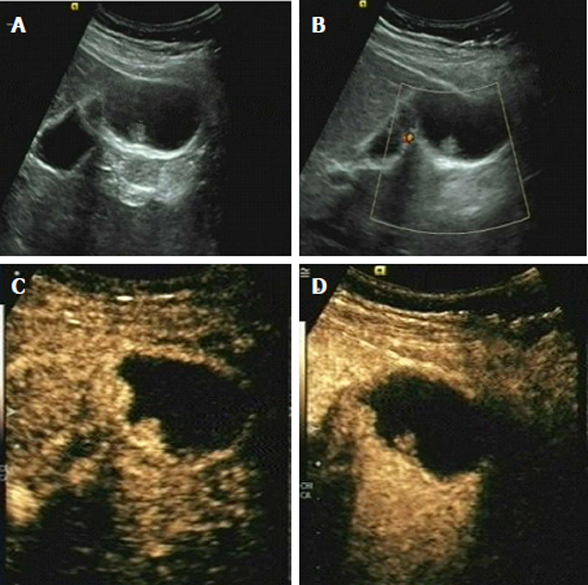
Figure 3 Gallbladder polyps in a 38-year-old male patient.
A: B-mode sonography showed a homogeneously isoechoic lesion in the gallbladder, with an intact gallbladder wall; B: Color Doppler ultrasound showed no color Doppler signal in the lesion. According to A and B, a diagnosis of probably benign was made. C: CEUS showed a homogeneous and a little hyperenhanced lesion in the gallbladder on arterial phase; D: CEUS showed the enhancement of the lesion is similar to the surrounding gallbladder wall on venous phase. According to C and D, a diagnosis of benign lesion was made. CEUS: Contrast-enhanced ultrasound.
- Citation: Zhang HP, Bai M, Gu JY, He YQ, Qiao XH, Du LF. Value of contrast-enhanced ultrasound in the differential diagnosis of gallbladder lesion. World J Gastroenterol 2018; 24(6): 744-751
- URL: https://www.wjgnet.com/1007-9327/full/v24/i6/744.htm
- DOI: https://dx.doi.org/10.3748/wjg.v24.i6.744


