Published online Mar 14, 2007. doi: 10.3748/wjg.v13.i10.1628
Revised: February 15, 2006
Accepted: March 6, 2007
Published online: March 14, 2007
Gynura root has been used extensively in Chinese folk medicine and plays a role in promoting microcirculation and relieving pain. However, its hepatic toxicity should not be neglected. Recently, we admitted a 62-year old female who developed hepatic veno-occlusive disease (HVOD) after ingestion of Gynura root. Only a few articles on HVOD induced by Gynura root have been reported in the literature. It is suspected that pyrrolizidine alkaloids in Gynura root might be responsible for HVOD. In this paper, we report a case of HVOD and review the literature.
- Citation: Dai N, Yu YC, Ren TH, Wu JG, Jiang Y, Shen LG, Zhang J. Gynura root induces hepatic veno-occlusive disease: A case report and review of the literature. World J Gastroenterol 2007; 13(10): 1628-1631
- URL: https://www.wjgnet.com/1007-9327/full/v13/i10/1628.htm
- DOI: https://dx.doi.org/10.3748/wjg.v13.i10.1628
Hepatic veno-occlusive disease (HVOD) is a clinical syndrome characterized by hyperbilirubinemia, painful hepatomegaly and weight gain due to fluid retention, after hematopoietic stem cell transplantation (HSCT), HVOD is a well-recognized life threatening complication, with an incidence rate of 10% to 60%[1]. In 1920, Willmot and Robertson[2] reported that HVOD is associated with the ingestion of Senecio tea, which contains pyrrolizidine alkaloids (PA). Other herb or plant medicine containing PA has been reported to cause hepatic injury and hepatic sinusoidal-obstruction syndrome[3]. A few cases of HVOD relating to Gynura root usage have been reported in Chinese literature[4-9]. Gynura root has been used extensively in Chinese folk medicine and plays a role in promoting microcirculation and relieving pain and curing injury.
Recently, we admitted a 62-year old female who presented with ascites, abdominal distention and was finally confirmed to have HVOD. This patient had a history of ingestion of Gynura root and experienced series of diagnostic approaches and various therapies. Since Gynura root or its analog may be used elsewhere in the world, we report the case and review the literature.
A 62-year old woman was admitted on January 23, 2006 to Hepato-gastroenterology Department of our hospital with abdominal complaints. She had abdominal distention after eating for about 10 mo. An abdominal ultrasound examination at a local hospital showed no remarkable findings. She had upper abdominal pain and weight gain in recent 3 mo. One week ago, abdominal pain became more severe, and repeated ultrasound examination revealed hepatomegaly and ascites. She had no fever and night sweats, no contact with sick persons or animals. Past medical history revealed asthma for over 20 years, which was treated occasionally with inhaler. She had no history of liver disease, alcohol abuse, pulmonary tuberculosis (TB). Family history was not significant.
Her initial vital signs were normal. Physical examination showed abdominal distention with upper abdominal tenderness, mildly dilated superficial abdominal veins, hepatomegaly (liver 7 cm below the xiphoid process), but no palpable spleen. Lower extremities showed no peripheral edema.
Laboratory tests showed normal hemoglobin (13.7 g/dL), normal WBC count (4700/μL), normal differentiatiation, but low platelets of 75 000/μL. Serum total protein (62.1 g/L) and albumin (31.9 g/L) were slightly below normal (protein normal range 63-82 g/L, Albumin 35-53 g/L). Other liver function tests showed normal total bilirubin, direct bilirubin, alanine aminotransferase and gammaglutamyl transferase, but slightly elevated aspartate aminotransferase 38 U/L (normal 5-35 U/L) and alkaline phosphatase 122 U/L (normal 30-110 U/L). Prothrombin time was normal and hepatitis B/C serology was negative. Serum tumor markers of alpha-fetoprotein (AFP), carcinoembryonic antigen (CEA) and CA-199 were all normal but cancer antigen 125 (CA-125) was elevated (49.87 U/mL, normal range < 35 U/mL). Examination of ascetic fluid showed that the fluid was like exudate and transudate in appearance. The albumin level was elevated (20 g/L). The serum-ascites albumin gradient (SAAG) was 11.9 g/L. The ascitic cytology was negative for malignancy. Ziehl-Neelsen stain of the ascites was negative for TB. PPD test was negative.
An abdominal computerized tomography (CT) revealed hepatomegaly and residual ascites. No hepatic veins were visualized, suggesting that there was obstruction of hepatic vein outflow. No abnormality was seen in the pancreas, spleen, mesentery, retroperitoneal and pelvic organs. A digital subtraction angiography (DSA) showed normal left and right hepatic veins. There was no stricture or obstruction of the inferior vena cava, hepatic artery and portal vein. The patient was treated symptomatically with fluid restriction, diuretics,and albumin.
On her 23rd day of hospitalization (Feb 14), because of lack of clinical improvement and uncertainty of her diagnosis, exploratory laparoscopy was performed. At operation, about 3000 mL ascites was removed. Pelvic organs (uterus, ovaries and tubes), small and large intestines, stomach, omentum and diaphragm were normal. The liver appeared diffusely congested and the left lobe lateral segment was enlarged. Portal venous pressure of about 24 mmHg was measured in a branch of the mesenteric vein. Two biopsies of the liver were taken and histology showed that the central and sublobular veins and hepatic sinus were prominently congested and dilated. Hepatic cells appeared atrophic and degenerated. The walls of sublobular veins were thickened, and the hepatic venules revealed significant fibrosis. Based on the clinical manifestations and these histological findings, HVOD was confirmed.
One week later, because of increased abdominal distension and edema of lower legs and perineum, contrast enhanced timing robust angiography (ceMRA) was done showing narrowing of hepatic veins, uneven distribution of contrast material in hepatic parenchyma, suggesting venous congestion. The inferior vena cava, portal vein, abdominal aorta, and bilateral renal arteries appeared to be normal.
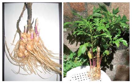
She had ingested Gynura root (tu san qi) for 3 mo before admission to our hospital for neck pain secondary to cervical osteophyte. She was treated with 3 slices of fresh Gynura root a day (about 2 g/d) soaked in rice wine in a bowl. Then she placed the bowl in a pot with water and steamed it. She ate the Gynura root and drank the wine. During the three months, she did not receive any Western medication and any other herbal remedy. We planted the Gynura root till it was full grown (Figure 1). The plant was identified as gynura segetum.
The patient was then treated with low molecular weight heparin and prostaglandin E1 and diuretics, etc, but her symptoms did not improve. Since methylprednisolone was tried for three weeks without much better effect, a transjugular intrahepatic portosystemic shunt (TIPS) was attempted on Mar 17. The puncture wire and needle could be passed through the hepatic veins into the portal vein branches. However, the stent could not be inserted because of the narrowing portal branches. TIPS failed. After a series of supportive therapy and consultations, the patient was transferred to the Surgery Department to undergo laparotomy and a mesocaval shunt was performed on Mar 29, 2006. At operation, the liver was found to be diffusely congested, the superior mesenteric vein was connected to the inferior vena cava with a 10 mm Gore-Tex graft. Initially, mesenteric venous pressure was 26 mm Hg. After the shunt was in place, the venous pressure decreased to 17 mmHg. After surgery, the patient received treatment with antibiotics, anticoagulant and tapering of her steroids. Anticoagulant was started from the 3rd post-operative day for 5 d, oral medication for two weeks, then subcutaneously for two more weeks. Two weeks post-operation, the patient developed pneumonia, treated and was discharged one month later. During follow-up examination in June of 2006, she had no abdominal distention nor pain. Repeated abdominal ultrasound study showed no ascites and the liver size was normal.
Since Gynura root is widely used in Chinese folk medicine, we searched the main Chinese medical database and found 6 Chinese publications with 21 cases of Gynura root-related HVOD[4-9], 11 of them were confirmed by liver biopsy or autopsy. The clinical characteristics of the Gynura root-related HVOD patients are summarized in Table 1. All the patients presented with hepatomegaly and ascites. Two patients died after ingestion of high doses of Gynura root (450 g and 1800 g within 10 d and 50 d respectively)[4]. Most of the patients had elevated total bilirubin, and only a few cases had a normal total bilirubin.
| Reference | Sex | Age(yr) | Reason for useof gynura root | duration | Total gynura(gram) | Total bilirubin(nl range4) | Special testsof HVOD | Therapy forHVOD | Outcome |
| Hou JG, 1980[4] | M | 57 | Coronary heart disease | About 10 d | 450 | Normal | Autopsy | Symptomatic | Died 5-d after admission |
| F | 48 | Diabetes | About 50 d | 1800 | 4.5 mg% | Ultrasound, liver scan, autopsy | Symptomatic | Died | |
| Li JP 2001[5] | M | 53 | No detail | No detail | No detail | 53.1 μmol/L (0-20.4) | Ultrasound, MRI, Biopsy | In situ liver transplantation | Well |
| Li ZM, 2005[6] | M | 43 | Back ache | 20 d | 150 | No report | Ultrasound MRI, DSA, Biopsy | Methylprednisolone 40 mg IV q 12 h | Well |
| Chen WX, 2005[7] | M | 52 | After a fall | 4 mo | 3601 | 27 μmol/L (0-20.4) | Ultrasound, CT, Biopsy | In situ liver transplantation | Well 45-d after operation |
| F | 39 | Trauma | 3 mo | 1802 | 25 μmol/L (0-20.4) | Ultrasound CT, Biopsy | Symptomatic treatment | Discharged without improvement | |
| Yan Hong 2005[8] | M | 29 | nephrolith | 20 d | 200 | No report | Ultrasound, CT, DSA, Biopsy | Prednisone | Well |
| Our case | F | 62 | Neck pain | 3 mo | 1803 | 17 μmol/L (0-20.4) | Ultrasound, CT, DSA, Biopsy, ceMRA | Mesocaval shunt | Well 2-mo later |
| Zhang GH 2006[9] | M6, F8 | 41-73 | Trauma or fracture | 5 d (mean) | 300-700 | Elevated in 12 cases | All had ultrasound, CT; 4 cases biopsy; 2 cases DSA | Low molecular weight heparin, aspirin and diuretics | Well |
HVOD which was first described in 1920 by Willmot and Robertson[2] is associated with the ingestion of Senecio tea containing pyrrolizidine alkaloids (PA). In 1953, Hill et al[10] reported a large series of 150 Jamaican children who developed hepatic HVOD after drinking Senecio tea. More than 300 kinds of PA have been identified in over 6000 plants of the Compositae, Boraginaceae and Leguminosae families[3]. Gynura root belongs to the Compositae family.
Yuan et al[11] isolated six alkaloids from Gynura segetum (Lour.) Merr. Two of the six were identified as senecionine and seneciphylline, which are known to have hepatic toxicity. These PAs, which have minimal toxicity in their original form, are metabolized in the liver through CYP (P450 cytochrome) and become toxic metabolites. The latter can act on local liver cells to cause damage by cross-linking DNA[12,13]. Furthermore, PA can decrease glutathione (GSH) in sinusoidal endothelial cells[14]. This enhanced oxidative stress also can affect collagen α1 transcription directly and/or through the activation of hepatic stellate cells[15], finally leading to HVOD. If the liver becomes damaged, the pyrrolizidine metabolites can overflow and infiltrate the lung fluids and cause damage there. Pulmonary edema and pleural effusions may occur, sometimes resulting in fatalities with very high levels of PA ingestion[13].
Confirmation of HVOD is based on the histology examination of liver tissue. The hepatic sinus, central and sublobular vein are significantly congested and dilated in Gynura root-related HVOD, while the venular walls are thickened with collagen deposition, with or without infiltration of lymphocytes, monocytes and neutrophils with fatty degeneration and necrosis presented in hepatic cells[4-9].
Currently, various strategies are used for the treatment of HVOD. For herb-related HVOD, plants containing PA should be avoided and discontinued. Some patients may recover after symptomatic treatment such as fluid restriction, diuretics and albumin. Administration of methylprednisolone seems to be another favorable alternative for some HVOD patients[6,16]. For patients with serious HVOD who do not respond to medical therapy, early use of transjugular intrahepatic portosystemic shunt (TIPS) can be considered[17]. However, it was reported that TIPS should not recommended for patients with HVOD[17,18]. In our patient, TIPS was not performed and she recovered well after undergoing meso-caval shunt. The value of porto-systemic shunt in HVOD remains to be further investigated. For those critically ill patients without response to porto-systemic shunt, liver transplantation might be considered with a survival rate of about 30%[19]. Li et al[6] and Chen et al[7] reported that two male cases of Gynura-root related HVOD recovered well over 40 d after liver transplant for hepatic failure.
Some researchers have suggested that PAs related safety problems are more widespread in certain areas of the world, such as Africa and South America[13]. Gynura root-related HVOD may be more widespread in China as well. Eighteen of these 22 patients (including our patient) were residents of Zhejiang Province, eastern coastal China, where more concerns are paid on Gynura root-related HVOD through organized case discussions between hospitals. Supervision and instruction of PA content in herbal medicine are crucial. The German Health Administration has set a standard for the use of herb petasites[13]. Further measures should be taken to determine and supervise herbs containing PAs to reduce their toxic effect, and make plentiful use of the Chinese traditional herbal medicine.
The authors thank Yeu-Tsu Margaret Lee, MD, FACS, Department of Surgery, School of Medicine, University of Hawaii, USA and professor Pe-Hsun Tung, Department of Surgery, Washington County Hospital for their assistance in preparing this paper. We also thank Professor You-Fa Zhu, Department of Pathology, College of Medicine, Zhejiang University for his technical support in the histological examination.
S- Editor Liu Y L- Editor Wang XL E- Editor Lu W
| 1. | McDonald GB, Sharma P, Matthews DE, Shulman HM, Thomas ED. Venocclusive disease of the liver after bone marrow transplantation: diagnosis, incidence, and predisposing factors. Hepatology. 1984;4:116-122. [RCA] [PubMed] [DOI] [Full Text] [Cited by in Crossref: 616] [Cited by in RCA: 582] [Article Influence: 14.2] [Reference Citation Analysis (0)] |
| 2. | Willmot FC, Robertson GW. Senecio disease, or cirrhosis of the liver due to Senecio poisoning. Lancet. 1920;2:848. [RCA] [DOI] [Full Text] [Cited by in Crossref: 141] [Cited by in RCA: 117] [Article Influence: 1.1] [Reference Citation Analysis (0)] |
| 3. | Chojkier M. Hepatic sinusoidal-obstruction syndrome: toxicity of pyrrolizidine alkaloids. J Hepatol. 2003;39:437-446. [RCA] [PubMed] [DOI] [Full Text] [Cited by in Crossref: 100] [Cited by in RCA: 90] [Article Influence: 4.1] [Reference Citation Analysis (0)] |
| 4. | Hou JG, Xia YT, Yu CS, An R, Tang YH. Hepatic veno-occlusive disease: report of two cases. Chin J Intern Med. 1980;3:187-191. |
| 5. | Li JP, Hu MH, Jin HH, Dai T, Zhu LF. Orthotopic liver transplantation in treating hepatic veno-occlusive disease. Zhongguo Gandan Waike Zazhi. 2001;7:inside front cover. |
| 6. | Li ZM, Pan WS, Cai J T, Chen L R, Yao M. Successful treatment of hepatic veno-occlusive disease caused by gynura root-a case report. Zhonghua Neike Zazhi. 2005;44:144-145. |
| 7. | Chen WX, Yang M, Yu CH, Li YM. Hepatic veno-occlusive disease: report of two cases. Zhonghua GanZangBing ZaZhi. 2005;13:394-395. [PubMed] |
| 8. | Yan H, Bai L, Peng M. Ju san qi caused acute hepatic toxicity: one case report. Zhonghua Ganzhangbing Zazhi. 2005;10:344. |
| 9. | Zhang GH, Kong AZ, Fang JW, Chen YJ, Zheng WL, Dong DJ, Zhang SZ. CT imaging of hepatic veno-occlusive disease (an analysis of 14 cases). Linchuang Fangshexue Zazhi. 2006;13:250-254. |
| 10. | Hill KR, Rhodes K, Stafford JL, Aub R. Serous hepatosis: a pathogenesis of hepatic fibrosis in Jamaican children. Br Med J. 1953;1:117-122. [RCA] [PubMed] [DOI] [Full Text] [Cited by in Crossref: 40] [Cited by in RCA: 31] [Article Influence: 0.4] [Reference Citation Analysis (0)] |
| 11. | Yuan SQ, Gu GM, Wei TT. Studies on the alkaloids of Gynura segetum (Lour.) Merr. YaoXue XueBao. 1990;25:191-197. [PubMed] |
| 12. | Kim HY, Stermitz FR, Li JK, Coulombe RA. Comparative DNA cross-linking by activated pyrrolizidine alkaloids. Food Chem Toxicol. 1999;37:619-625. [RCA] [PubMed] [DOI] [Full Text] [Cited by in Crossref: 38] [Cited by in RCA: 31] [Article Influence: 1.2] [Reference Citation Analysis (0)] |
| 13. | Subhuti Dharmananda. Safety issues affecting herbs: pyrrolizidine alkaloids. Available from: http://www.itmonline.org/arts/pas.htm. |
| 14. | Wang X, Kanel GC, DeLeve LD. Support of sinusoidal endothelial cell glutathione prevents hepatic veno-occlusive disease in the rat. Hepatology. 2000;31:428-434. [RCA] [PubMed] [DOI] [Full Text] [Cited by in Crossref: 97] [Cited by in RCA: 94] [Article Influence: 3.8] [Reference Citation Analysis (0)] |
| 15. | Chojkier M, Houglum K, Lee KS, Buck M. Long- and short-term D-alpha-tocopherol supplementation inhibits liver collagen alpha1(I) gene expression. Am J Physiol. 1998;275:G1480-G1485. [PubMed] |
| 16. | Al Beihany A, Al Omar H, Sahovic E, Chaudhri N, Al Mohareb F, Al Sharif F, Al Zahrani H, Al Shanqeeti A, Seth P, Zaidi S. Successful treatment of hepatic veno-occlusive disease after myeloablative allogeneic hematopoietic stem cell transplantation by early administration of a short course of methylprednisolone. Bone Marrow Transplant. 2008;41:287-291. [PubMed] |
| 17. | Senzolo M, Cholongitas E, Patch D, Burroughs AK. TIPS for veno-occlusive disease: is the contraindication real? Hepatology. 2005;42:240-241; author reply 241. [RCA] [PubMed] [DOI] [Full Text] [Cited by in Crossref: 17] [Cited by in RCA: 19] [Article Influence: 1.0] [Reference Citation Analysis (0)] |
| 18. | Boyer TD, Haskal ZJ. The role of transjugular intrahepatic portosystemic shunt in the management of portal hypertension. Hepatology. 2005;41:386-400. [RCA] [PubMed] [DOI] [Full Text] [Cited by in Crossref: 315] [Cited by in RCA: 298] [Article Influence: 14.9] [Reference Citation Analysis (0)] |
| 19. | Kim ID, Egawa H, Marui Y, Kaihara S, Haga H, Lin YW, Kudoh K, Kiuchi T, Uemoto S, Tanaka K. A successful liver transplantation for refractory hepatic veno-occlusive disease originating from cord blood transplantation. Am J Transplant. 2002;2:796-800. [RCA] [PubMed] [DOI] [Full Text] [Cited by in Crossref: 28] [Cited by in RCA: 27] [Article Influence: 1.2] [Reference Citation Analysis (0)] |


