Copyright
©The Author(s) 2000.
World J Gastroenterol. Feb 15, 2000; 6(1): 102-106
Published online Feb 15, 2000. doi: 10.3748/wjg.v6.i1.102
Published online Feb 15, 2000. doi: 10.3748/wjg.v6.i1.102
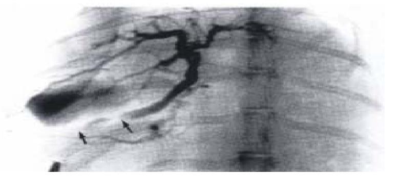
Figure 1 The cineradiographic recording of same bili ary tract of one control.
The contrast clustering the proximal segment (low-pre ssure ampulla) which enlarged significantly during excretion of contrast indicat ed that part was diastoled. (↑→↑: External segment of SO)
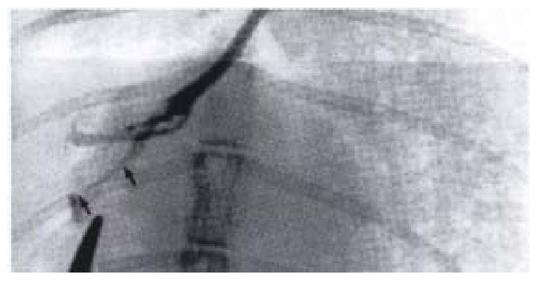
Figure 2 Biliary tract cineradiographic image of gr oup I rabbit.
The caliber of proximal segment decreased, retained a fixed spasm state. (↑→↑: External segment of SO)
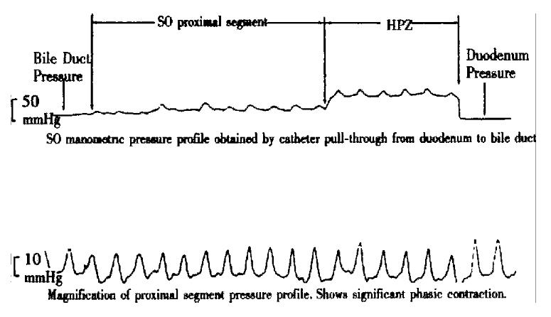
Figure 3 Pressure curve of SO segment of control rabbit.
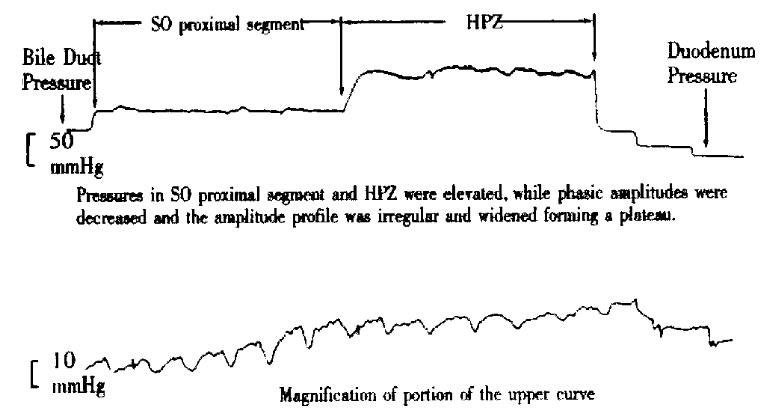
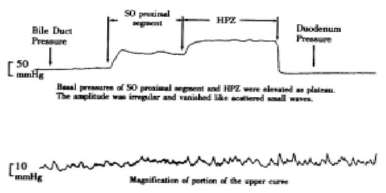
Figure 4 Pressure curve of SO segment of group I rabbit.
Figure 5 Pressure curve of SO segment of group II rabbit.
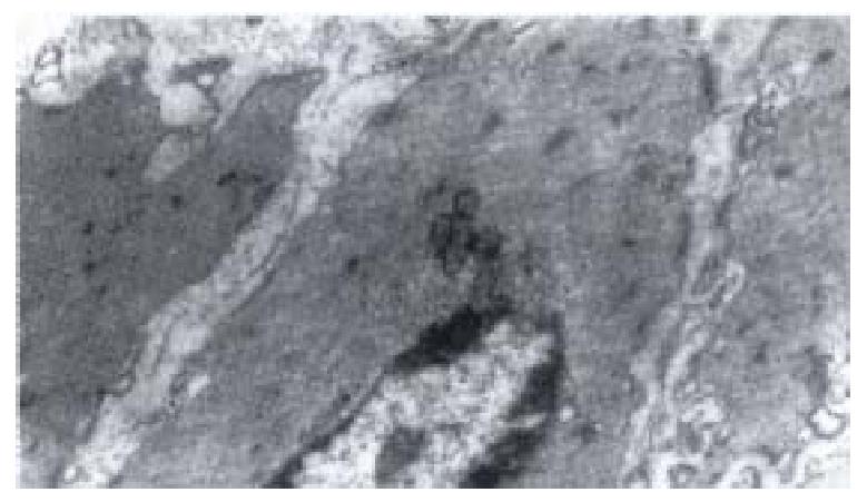
Figure 6 Electron-microscopic scanning of a control ‘s SO segment smooth muscle.
The myofilaments were regularly arranged, kink mac ula densa clear and dense, plasmosome was normal.
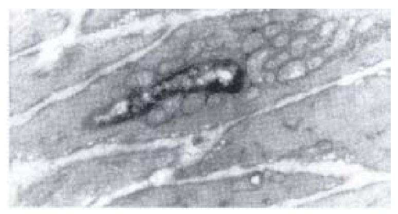
Figure 7 Electron-microscopic scanning of a group I rabbit’s SO smooth muscle.
Image showed the swelling of plasmosome and disap pear of intercristal space, decrease of kink macula densa and twisting of myofil aments.
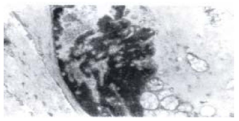
Figure 8 Electron-microscopic scanning of a group II rabbit’s SO segment smooth muscle.
It shows the swelling of plasmosome, irre gular arranged or fragmented intercristal space, some vacuolized, congregated at one end of nuclear, and disarragement of kink macula densa.
- Citation: Wei JG, Wang YC, Du F, Yu HJ. Dynamic and ultrastructural study of sphincter of Oddi in early-stage cholelithiasis in rabbits with hypercholesterolemia. World J Gastroenterol 2000; 6(1): 102-106
- URL: https://www.wjgnet.com/1007-9327/full/v6/i1/102.htm
- DOI: https://dx.doi.org/10.3748/wjg.v6.i1.102


