
















©The Author(s) 2000.
World J Gastroenterol. Feb 15, 2000; 6(1): 102-106
Published online Feb 15, 2000. doi: 10.3748/wjg.v6.i1.102
Published online Feb 15, 2000. doi: 10.3748/wjg.v6.i1.102
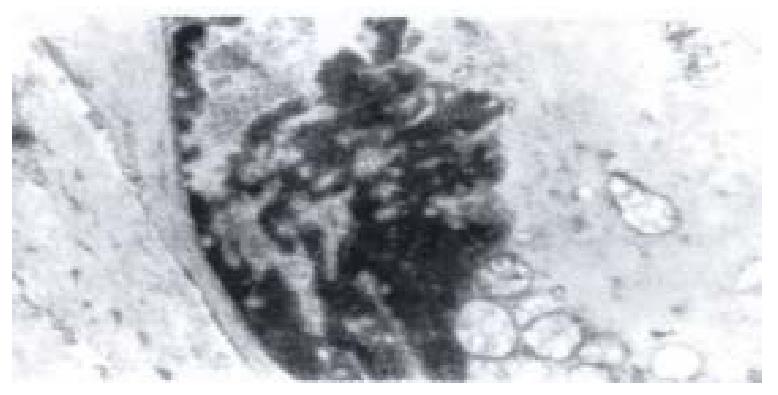
Figure 8 Electron-microscopic scanning of a group II rabbit’s SO segment smooth muscle.
It shows the swelling of plasmosome, irre gular arranged or fragmented intercristal space, some vacuolized, congregated at one end of nuclear, and disarragement of kink macula densa.
- Citation: Wei JG, Wang YC, Du F, Yu HJ. Dynamic and ultrastructural study of sphincter of Oddi in early-stage cholelithiasis in rabbits with hypercholesterolemia. World J Gastroenterol 2000; 6(1): 102-106
- URL: https://www.wjgnet.com/1007-9327/full/v6/i1/102.htm
- DOI: https://dx.doi.org/10.3748/wjg.v6.i1.102