
















Copyright: ©Author(s) 2026.
World J Gastroenterol. Dec 7, 2026; 32(45): 122826
Published online Dec 7, 2026. doi: 10.3748/wjg.122826
Published online Dec 7, 2026. doi: 10.3748/wjg.122826
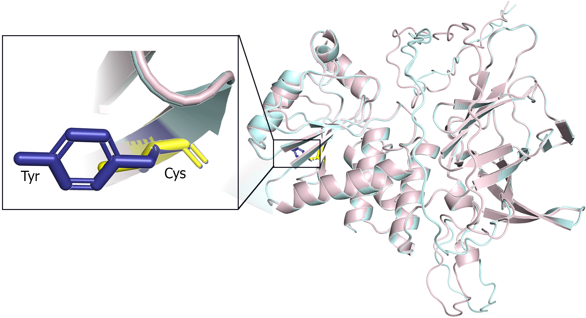
Figure 1 Three-dimensional structural model of the mutant PTEN protein.
α-helices are shown in light pink and β-sheets in light cyan. The inset provides a magnified view of the mutation site, displaying the spatial arrangement and side-chain conformations of the affected residues, with tyrosine (Tyr) shown in blue and cysteine (Cys) in yellow.
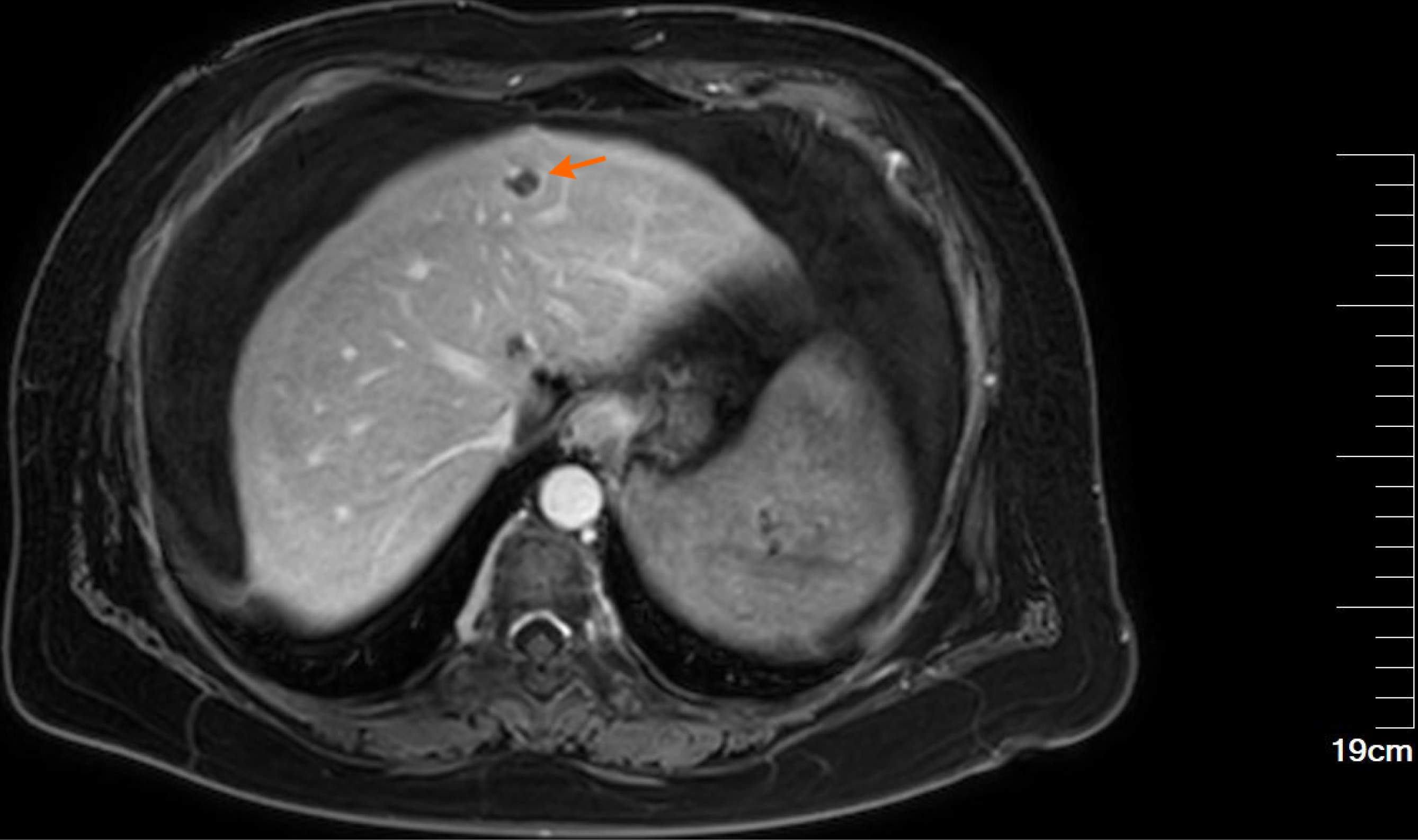
Figure 2 Upper abdominal magnetic resonance imaging.
The liver is slightly reduced in volume, with a wavy contour and widened fissures. A roundish lesion with hypointense T1-weighted and hyperintense T2-weighted signals is observed in the left hepatic lobe adjacent to the falciform ligament (orange arrow), measuring approximately 1.3 cm in diameter and demonstrating nodular enhancement on contrast-enhanced imaging. The portal vein is dilated, with a maximum diameter of 1.8 cm. Splenomegaly is present, with heterogeneous signal intensity and an irregular contour.
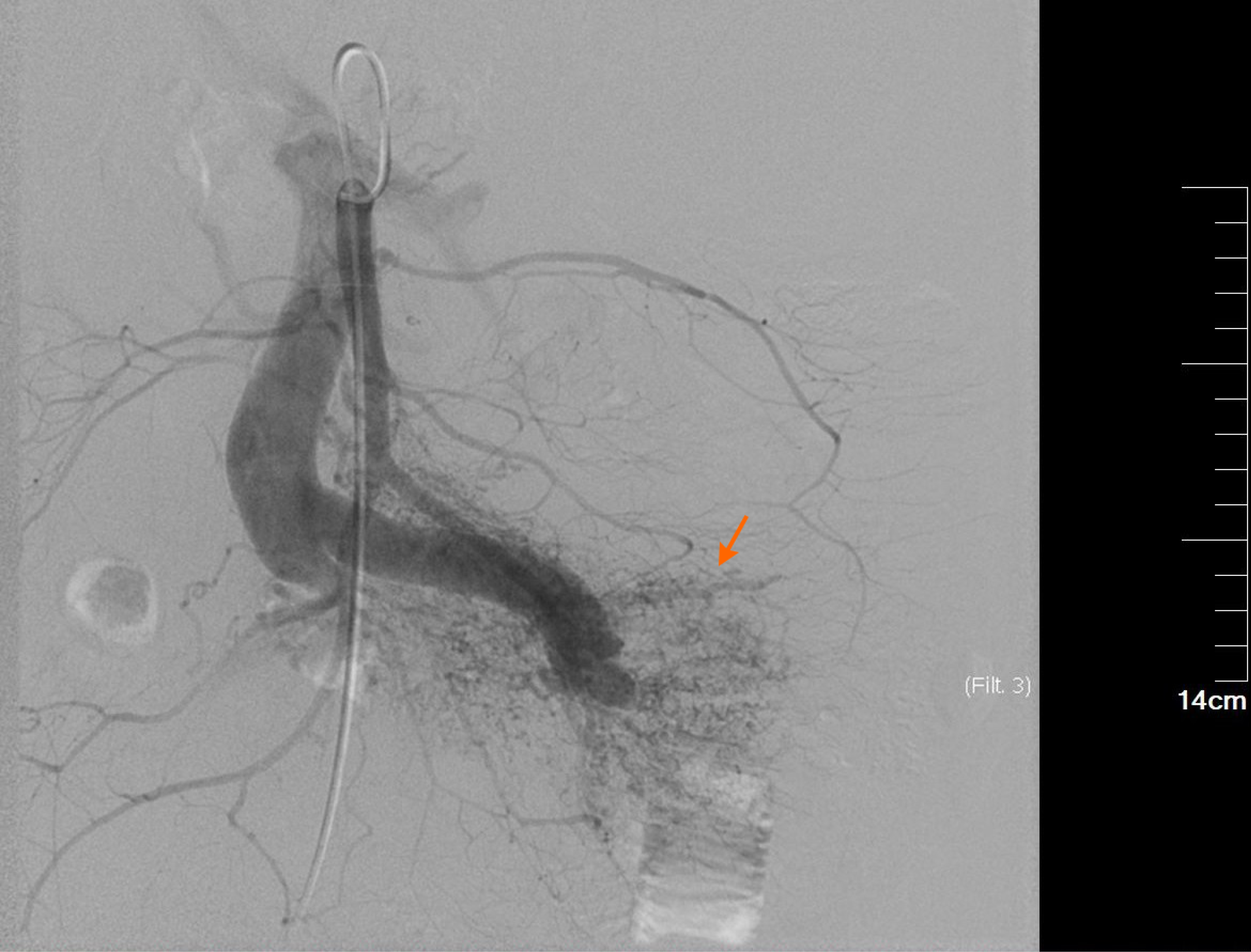
Figure 3 Digital subtraction angiography of the superior mesenteric artery with indirect portal venography.
The main trunk of the superior mesenteric artery appears thickened and tortuous. A distinct arteriovenous malformation is visible in the distal branches (orange arrow). No filling defects are observed within the vessels, and blood flow velocity is rapid, resulting in early visualization of the mesenteric and portal veins.
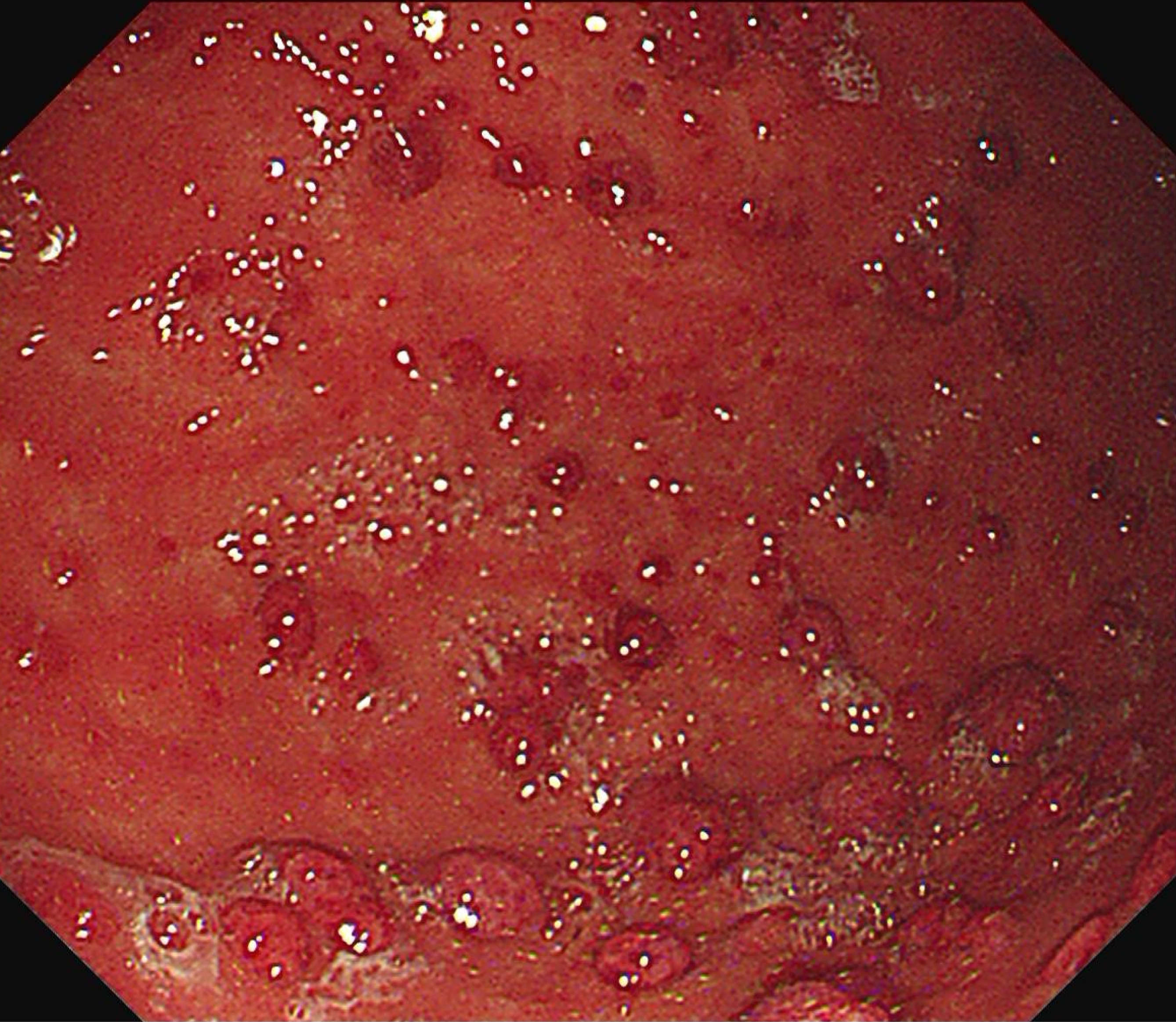
Figure 4 Upper gastrointestinal endoscopy.
Moderate turbid gastric fluid is present in the gastric body. Multiple scattered polypoid lesions measuring 0.5-1.2 cm in diameter were observed, with hyperemic surfaces. A biopsy specimen was obtained from a larger polyp located along the greater curvature. Histopathological examination confirmed a fundic gland polyp.
- Citation: Zhao ZH, Qi J, Hu JM, Zhao YY, Liu MQ, Deng L, Zhang F, Gao SS, Li K. Superior mesenteric arteriovenous fistula as a rare manifestation of Cowden syndrome: A case report and review of literature. World J Gastroenterol 2026; 32(45): 122826
- URL: https://www.wjgnet.com/1007-9327/full/v32/i45/122826.htm
- DOI: https://dx.doi.org/10.3748/wjg.122826