
















Copyright: ©Author(s) 2026.
World J Gastroenterol. Dec 7, 2026; 32(45): 122826
Published online Dec 7, 2026. doi: 10.3748/wjg.122826
Published online Dec 7, 2026. doi: 10.3748/wjg.122826
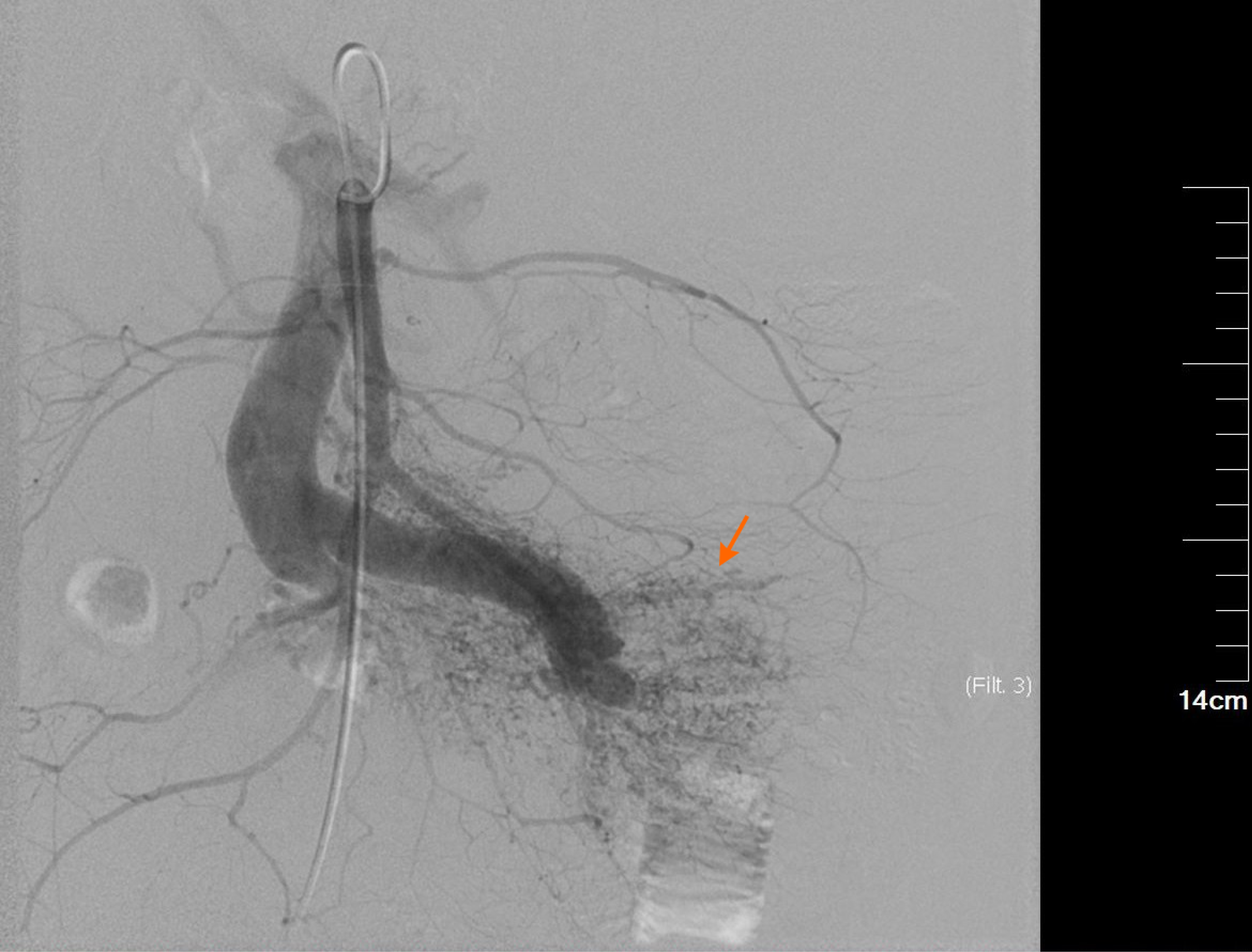
Figure 3 Digital subtraction angiography of the superior mesenteric artery with indirect portal venography.
The main trunk of the superior mesenteric artery appears thickened and tortuous. A distinct arteriovenous malformation is visible in the distal branches (orange arrow). No filling defects are observed within the vessels, and blood flow velocity is rapid, resulting in early visualization of the mesenteric and portal veins.
- Citation: Zhao ZH, Qi J, Hu JM, Zhao YY, Liu MQ, Deng L, Zhang F, Gao SS, Li K. Superior mesenteric arteriovenous fistula as a rare manifestation of Cowden syndrome: A case report and review of literature. World J Gastroenterol 2026; 32(45): 122826
- URL: https://www.wjgnet.com/1007-9327/full/v32/i45/122826.htm
- DOI: https://dx.doi.org/10.3748/wjg.122826