
















Copyright: ©Author(s) 2026.
World J Gastroenterol. Dec 7, 2026; 32(45): 122826
Published online Dec 7, 2026. doi: 10.3748/wjg.122826
Published online Dec 7, 2026. doi: 10.3748/wjg.122826
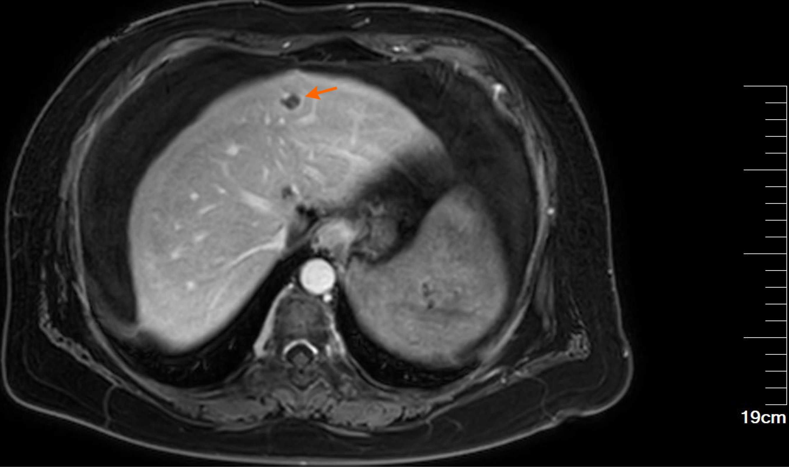
Figure 2 Upper abdominal magnetic resonance imaging.
The liver is slightly reduced in volume, with a wavy contour and widened fissures. A roundish lesion with hypointense T1-weighted and hyperintense T2-weighted signals is observed in the left hepatic lobe adjacent to the falciform ligament (orange arrow), measuring approximately 1.3 cm in diameter and demonstrating nodular enhancement on contrast-enhanced imaging. The portal vein is dilated, with a maximum diameter of 1.8 cm. Splenomegaly is present, with heterogeneous signal intensity and an irregular contour.
- Citation: Zhao ZH, Qi J, Hu JM, Zhao YY, Liu MQ, Deng L, Zhang F, Gao SS, Li K. Superior mesenteric arteriovenous fistula as a rare manifestation of Cowden syndrome: A case report and review of literature. World J Gastroenterol 2026; 32(45): 122826
- URL: https://www.wjgnet.com/1007-9327/full/v32/i45/122826.htm
- DOI: https://dx.doi.org/10.3748/wjg.122826