
















Copyright: ©Author(s) 2026.
World J Gastroenterol. Dec 7, 2026; 32(45): 122826
Published online Dec 7, 2026. doi: 10.3748/wjg.122826
Published online Dec 7, 2026. doi: 10.3748/wjg.122826
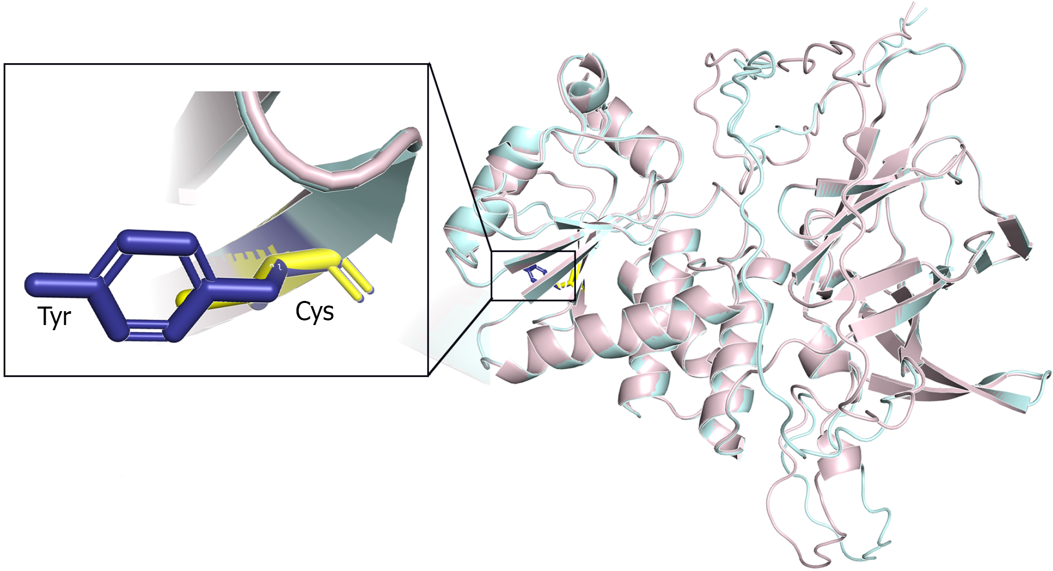
Figure 1 Three-dimensional structural model of the mutant PTEN protein.
α-helices are shown in light pink and β-sheets in light cyan. The inset provides a magnified view of the mutation site, displaying the spatial arrangement and side-chain conformations of the affected residues, with tyrosine (Tyr) shown in blue and cysteine (Cys) in yellow.
- Citation: Zhao ZH, Qi J, Hu JM, Zhao YY, Liu MQ, Deng L, Zhang F, Gao SS, Li K. Superior mesenteric arteriovenous fistula as a rare manifestation of Cowden syndrome: A case report and review of literature. World J Gastroenterol 2026; 32(45): 122826
- URL: https://www.wjgnet.com/1007-9327/full/v32/i45/122826.htm
- DOI: https://dx.doi.org/10.3748/wjg.122826