
















Copyright: ©Author(s) 2026.
World J Gastroenterol. Nov 28, 2026; 32(44): 120736
Published online Nov 28, 2026. doi: 10.3748/wjg.120736
Published online Nov 28, 2026. doi: 10.3748/wjg.120736
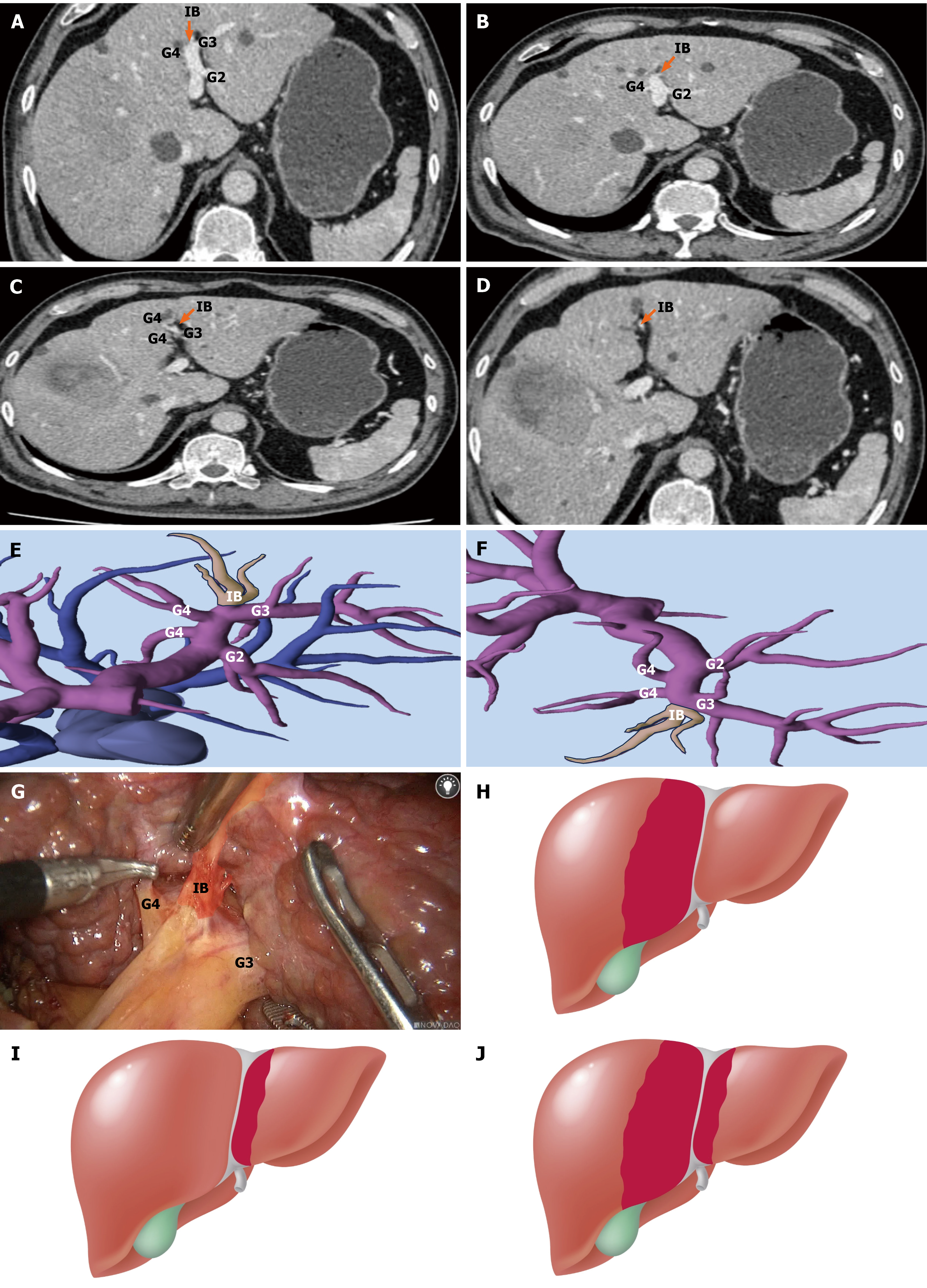
Figure 1 Portal territory-defined boundaries of segment 4 based on intermediate branches.
A-D: Axial contrast-enhanced computed tomography images showing intermediate branches (IBs; orange arrows) arising between the Glissonean pedicles of segments 3 and 4, supplying liver parenchyma on both sides of the falciform ligament; E and F: Three-dimensional reconstruction images demonstrating bilateral perfusion territories of the IBs, extending across the falciform ligament; G: Intraoperative laparoscopic view confirming the presence of IB during hepatic dissection; H: Schematic illustration of the traditional anatomical segment 4 (S4) boundary, with the left margin defined by the falciform ligament; I: Portal territory-based redefinition of the S4 territory showing leftward extension beyond the falciform ligament, in line with IB perfusion; J: Refined anatomical boundary of S4 according to the proposed portal vein-based classification, encompassing the full extent of perfused parenchyma. IB: Intermediate branch; G2: Glissonean pedicles of segment 2; G3: Glissonean pedicles of segment 3; G4: Glissonean pedicles of segment 4.
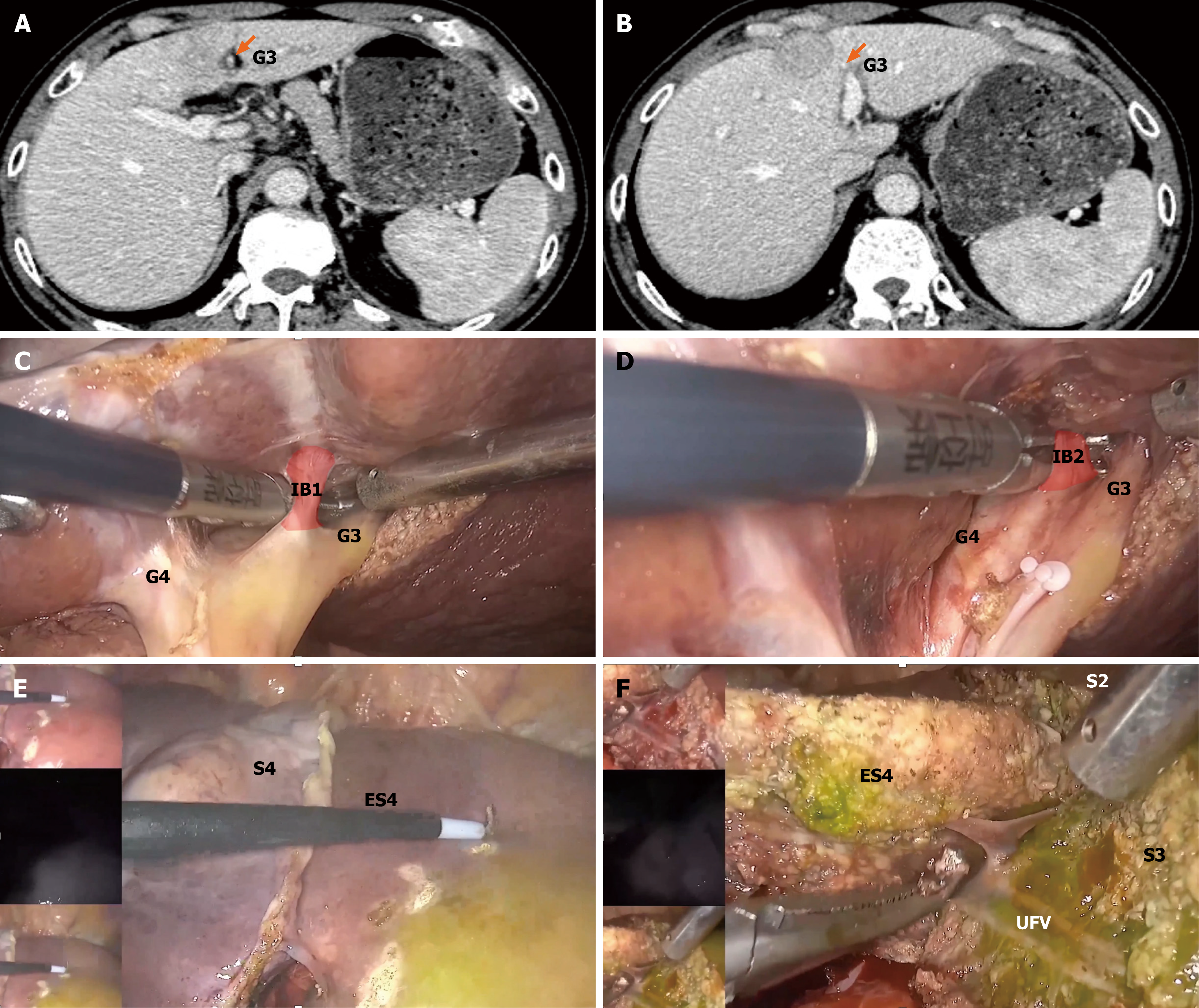
Figure 2 Portal territory-guided anatomical resection of extended segment 4.
A and B: Axial contrast-enhanced computed tomography images showing a lesion abutting the falciform ligament (orange arrows), a location that would typically require left hemihepatectomy under conventional anatomical definitions; C and D: Intraoperative laparoscopic views identifying two intermediate branches (IBs; IB1 and IB2) arising between the Glissonean pedicles of segments 3 and 4, supplying the parenchyma lateral to the falciform ligament; E: Indocyanine green (ICG) fluorescence negative staining delineating the extended segment 4 territory, corresponding to the perfusion zones of IB1 and IB2; F: Parenchymal transection guided by ICG staining and anatomical landmarks preserving segments 2 and 3, while achieving a negative surgical margin (R0). The umbilical fissure vein serves as a reference for the posterior limit of resection. IB: Intermediate branch; G3: Glissonean pedicles of segment 3; G4: Glissonean pedicles of segment 4; S2: Segment 2; S3: Segment 3; S4: Segment 4; ES4: Extended segment 4; UFV: Umbilical fissure vein serves.
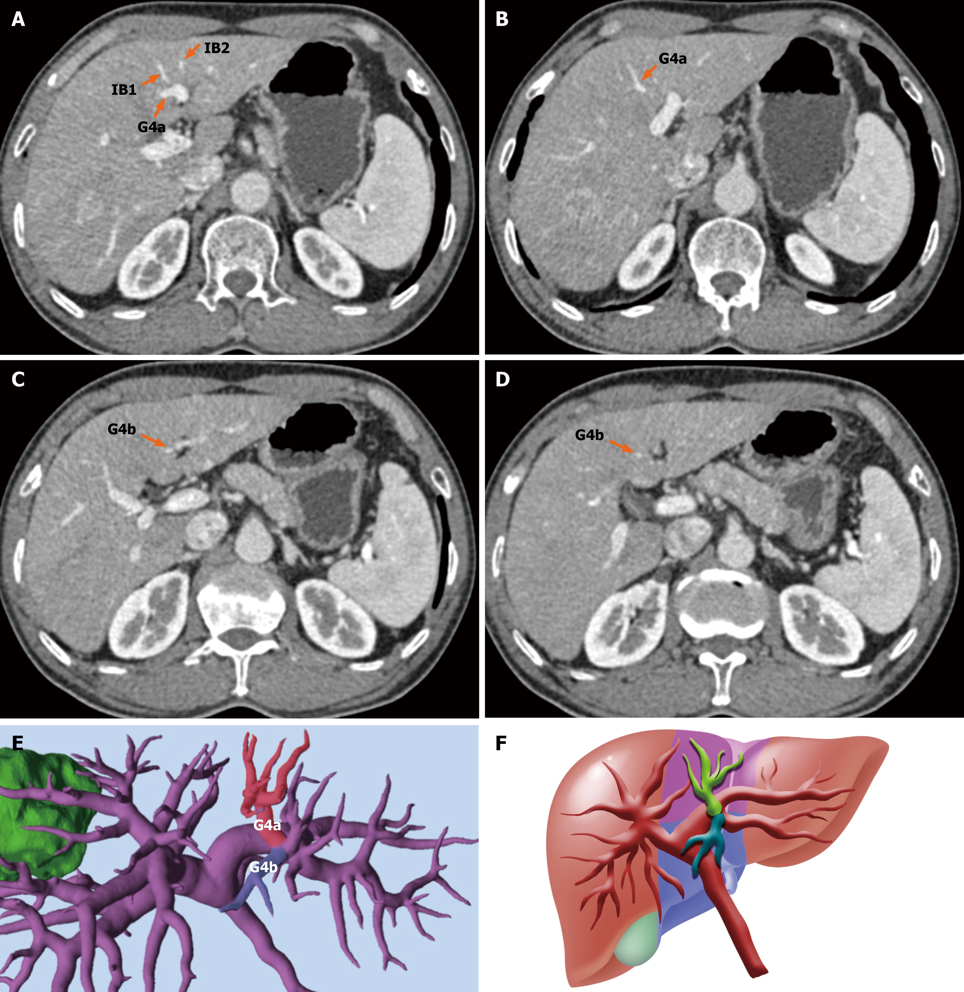
Figure 3 Type Ia classification: Bilateral cranial-caudal trunk type of segment 4.
A-D: Axial contrast-enhanced computed tomography images from a representative type Ia case, showing two dominant Glissonean trunks-Glissonean pedicles of segment 4 (G4; G4a and G4b)-supplying the cranial and caudal subsegments of segment 4 (S4), respectively. Two intermediate branches (IBs; IB1 and IB2) are also visible in panel A, arising near the bifurcation of segment 3 and S4; E: Three-dimensional portal vein reconstruction highlighting the bifurcated G4a and G4b pedicles supplying distinct cranial and caudal territories of S4; F: Schematic diagram illustrating the spatial distribution of G4a, G4b, and IBs, reinforcing the concept of bilateral cranial-caudal trunk anatomy in type Ia cases. IB: Intermediate branch; G4: Glissonean pedicles of segment 4.
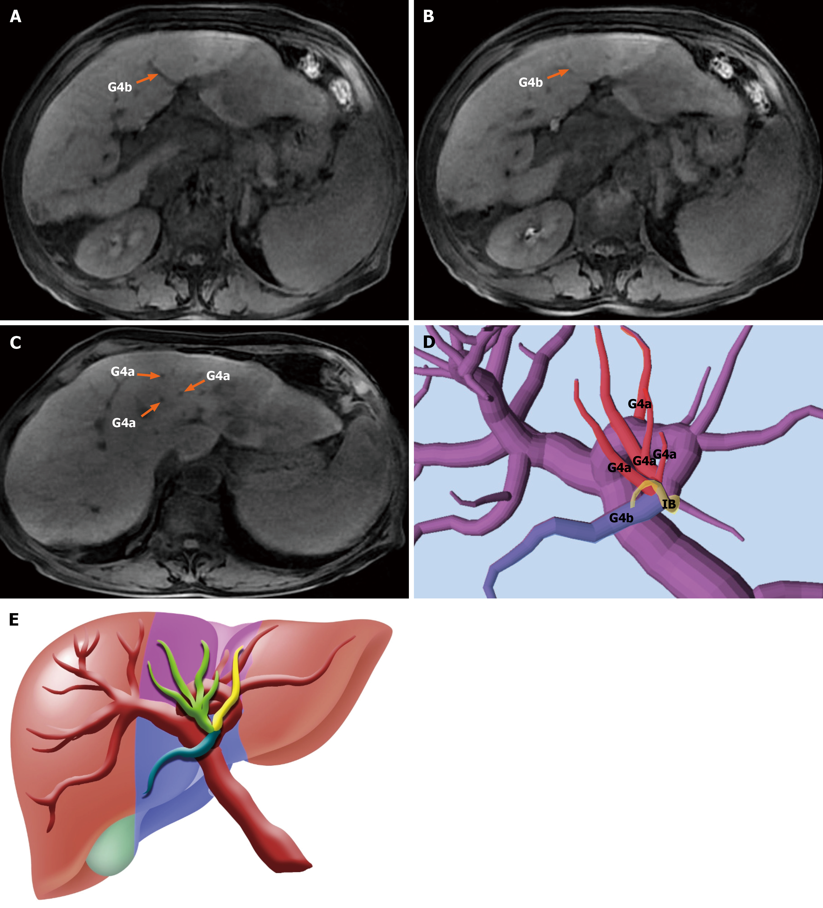
Figure 4 Type Ib classification: Unilateral cranial trunk with contralateral caudal dispersion in segment 4.
A-C: Gadolinium-enhanced magnetic resonance imaging images from a representative type Ib case. A dominant Glissonean pedicle (Glissonean pedicles of segment 4a) supplies the cranial portion of segment 4, while the caudal portion (Glissonean pedicles of segment 4b) receives scattered inflow from multiple smaller branches, indicating perfusion asymmetry; D: Three-dimensional reconstruction showing unilateral cranial inflow and contralateral caudal dispersion, with one intermediate branch arising near the bifurcation zone; E: Schematic diagram illustrating the type Ib pattern, with centralized cranial inflow and diffuse caudal distribution, reinforcing the concept of asymmetric cephalocaudal perfusion in this subtype. IB: Intermediate branch; G4: Glissonean pedicles of segment 4.
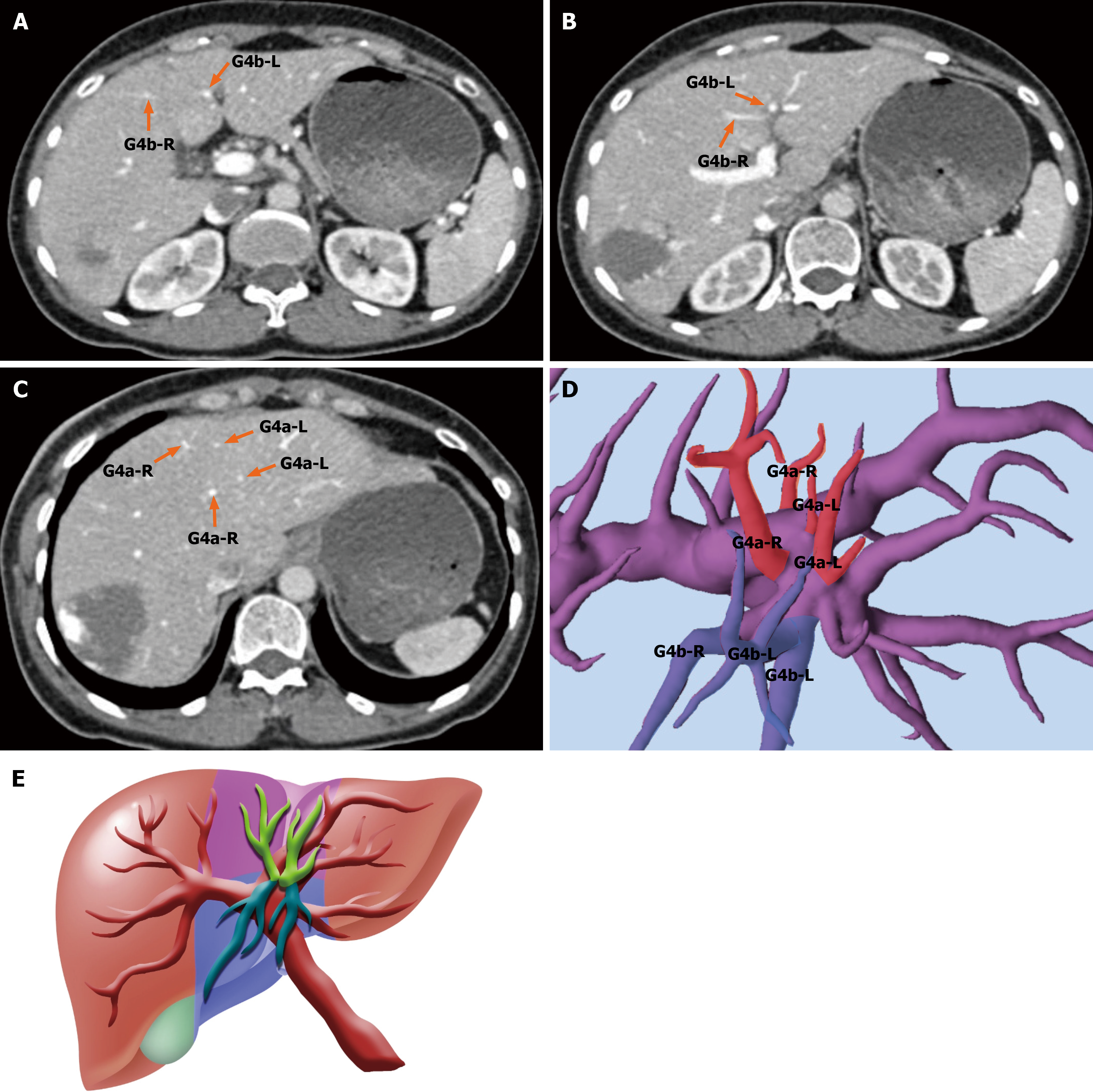
Figure 5 Type Ic classification: Fully dispersed Glissonean branching pattern in segment 4.
A-C: Axial contrast-enhanced computed tomography images from a representative type Ic case, showing fine and scattered Glissonean branches (Glissonean pedicles of segments 4a and 4b) supplying cranial and caudal subsegments without a dominant trunk; D: Three-dimensional portal vein reconstruction confirming a dispersed origin of multiple small Glissonean pedicles, each individually supplying subregions of segment 4 (S4); E: Schematic diagram illustrating the cranial-caudal perfusion pattern of S4 in type Ic, characterized by multiple independently arising Glissonean branches rather than a single dominant pedicle. G4: Glissonean pedicles of segment 4.
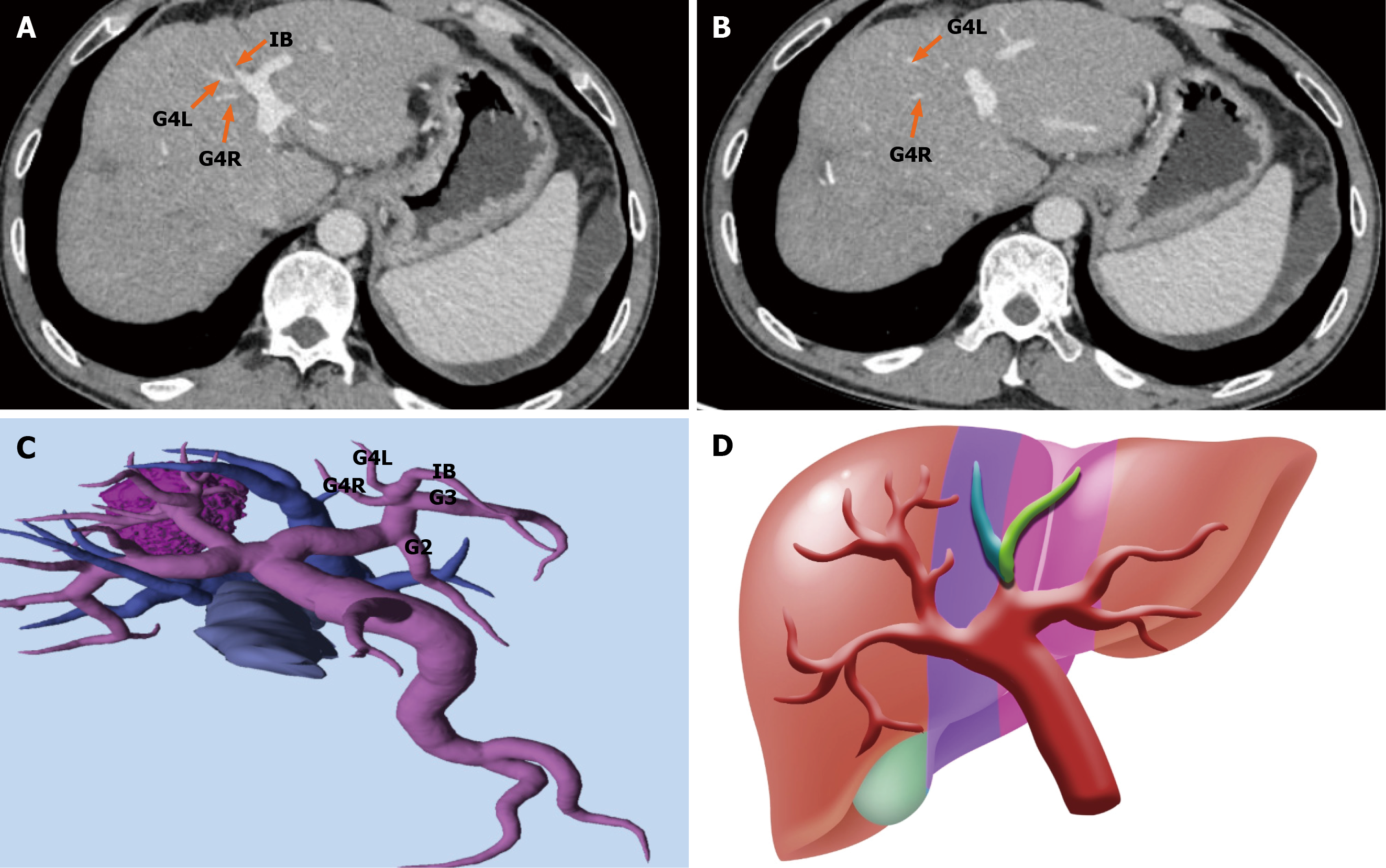
Figure 6 Type IIa classification: Bilateral trunk-type left-right subdivision of segment 4.
A and B: Axial contrast-enhanced computed tomography images from a representative type IIa case, showing two dominant Glissonean pedicles-Glissonean pedicles of segment 4 L (S4 L) and Glissonean pedicles of segment 4R (S4R)-supplying the left and right subsegments of S4 L and S4R, respectively; C: Three-dimensional portal vein reconstruction confirming a bilateral trunk-type configuration, with independent inflow to S4 L and S4R; D: Schematic diagram illustrating the symmetric left-right portal supply pattern, supporting lateralized subsegmental resection while preserving non-involved liver parenchyma. IB: Intermediate branch; G2: Glissonean pedicles of segment 2; G3: Glissonean pedicles of segment 3; G4: Glissonean pedicles of segment 4.
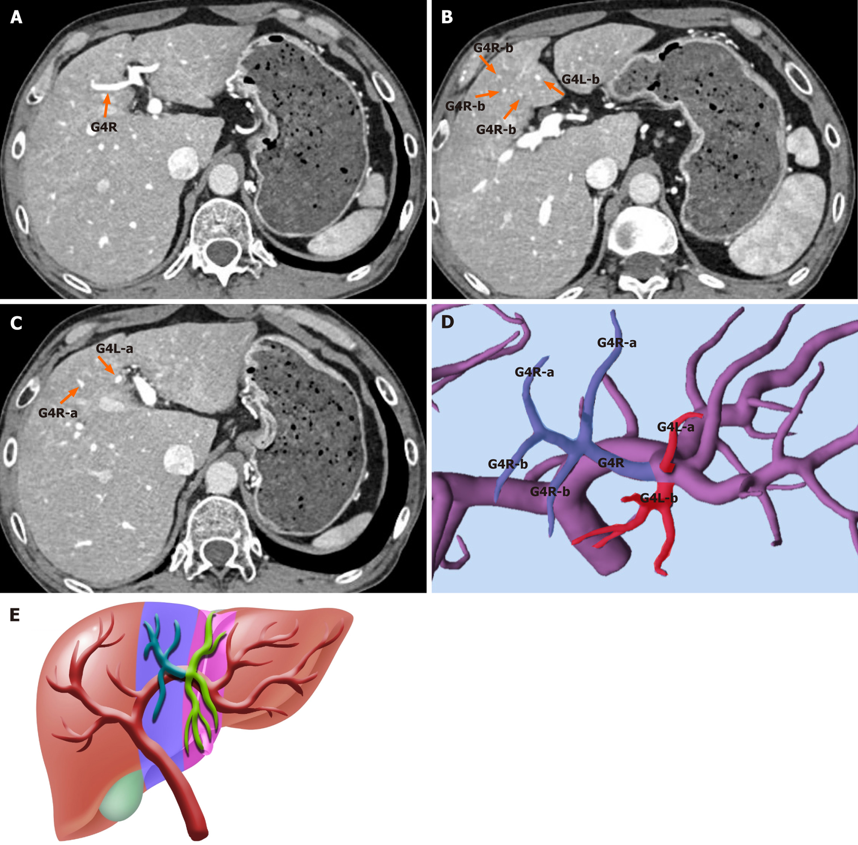
Figure 7 Type IIb classification: Unilateral trunk with contralateral dispersion in left-right subdivision of segment 4.
A-C: Axial contrast-enhanced computed tomography images from a representative type IIb case. A dominant Glissonean pedicles of segment 4 (G4; G4R) supplies the right subsegment of segment 4 (S4; S4R), while the left subsegment (S4 L) receives multiple fine and scattered branches (G4 L), without a dominant trunk; D: Three-dimensional portal vein reconstruction confirming the asymmetric left-right perfusion pattern, with a single structured G4R pedicle and dispersed G4 L branches; E: Schematic diagram illustrating the type IIb anatomy, characterized by unilateral trunk-type inflow to S4R and contralateral scattered inflow to S4 L. G4: Glissonean pedicles of segment 4.
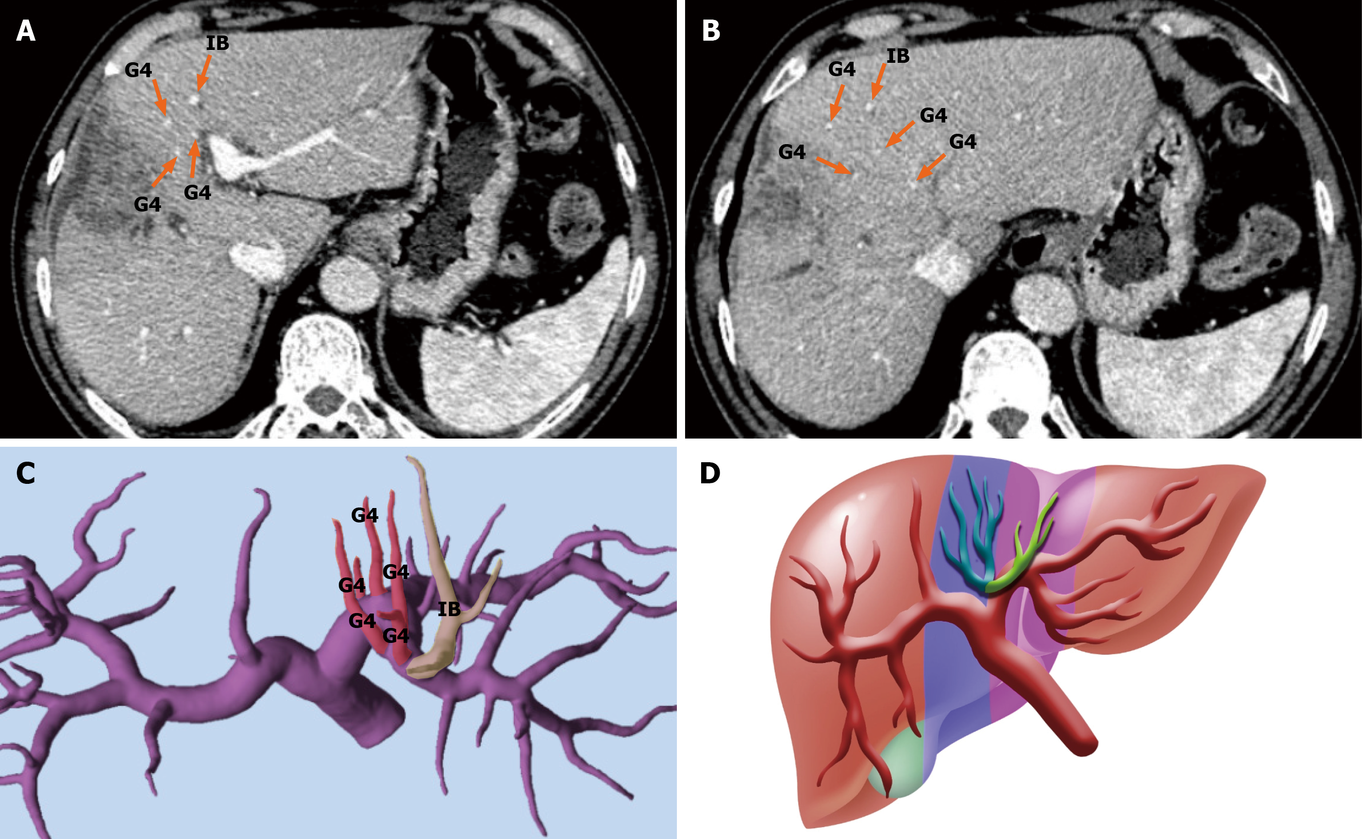
Figure 8 Type III classification: Scattered Glissonean inflow without dominant trunk anatomy in segment 4.
A and B: Axial contrast-enhanced computed tomography images from a representative type III case, showing multiple small Glissonean branches entering segment 4 (S4) from varying directions, with no discernible cranial-caudal or left-right segmentation pattern; C: Three-dimensional portal vein reconstruction revealing a disorganized inflow configuration, composed of scattered pedicles and intermediate branches supplying overlapping territories within S4; D: Schematic diagram illustrating the non-converging, diffuse vascular distribution in type III, which lacks structured segmentation and requires intraoperative fluorescence guidance for accurate subsegmental resection. IB: Intermediate branch; G4: Glissonean pedicles of segment 4.
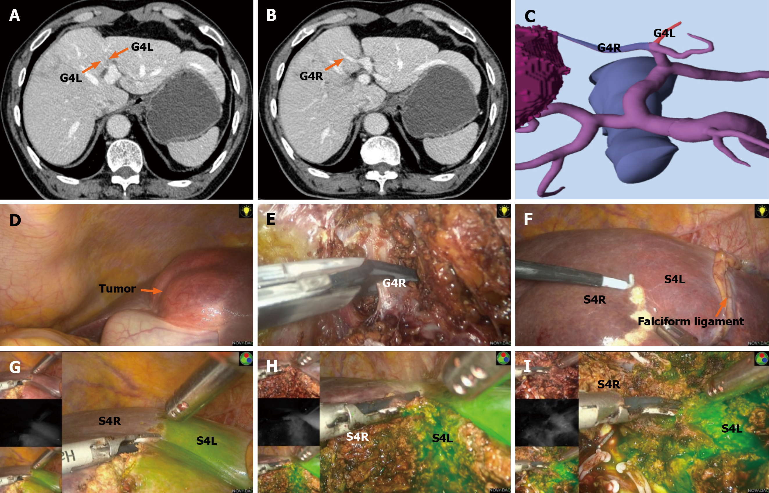
Figure 9 Portal-based subsegmental resection of segment 4 L and segment 4R guided by Glissonean anatomy and indocyanine green fluorescence demarcation.
A and B: Axial contrast-enhanced computed tomography images demonstrating separate inflow from the Glissonean pedicles of segment 4 (G4; G4 L and G4R) pedicles to the left and right of segment 4 (S4; S4 L and S4F) subsegments of S4, respectively (orange arrows); C: Three-dimensional portal vein reconstruction confirming a bilateral trunk-type configuration supplying S4 L and S4R; D and E: Intraoperative views showing identification and isolation of the G4R pedicle using the Glissonean approach; F: Surface marking of the liver based on anatomical landmarks delineating the boundary between S4 L and S4R; G-I: Indocyanine green fluorescence negative staining revealing distinct perfusion territories of S4 L and S4R, enabling precise subsegmental resection with functional parenchymal preservation and negative margins (R0). See Video 2 for operative details. S4: Segment 4; G4: Glissonean pedicles of segment 4.
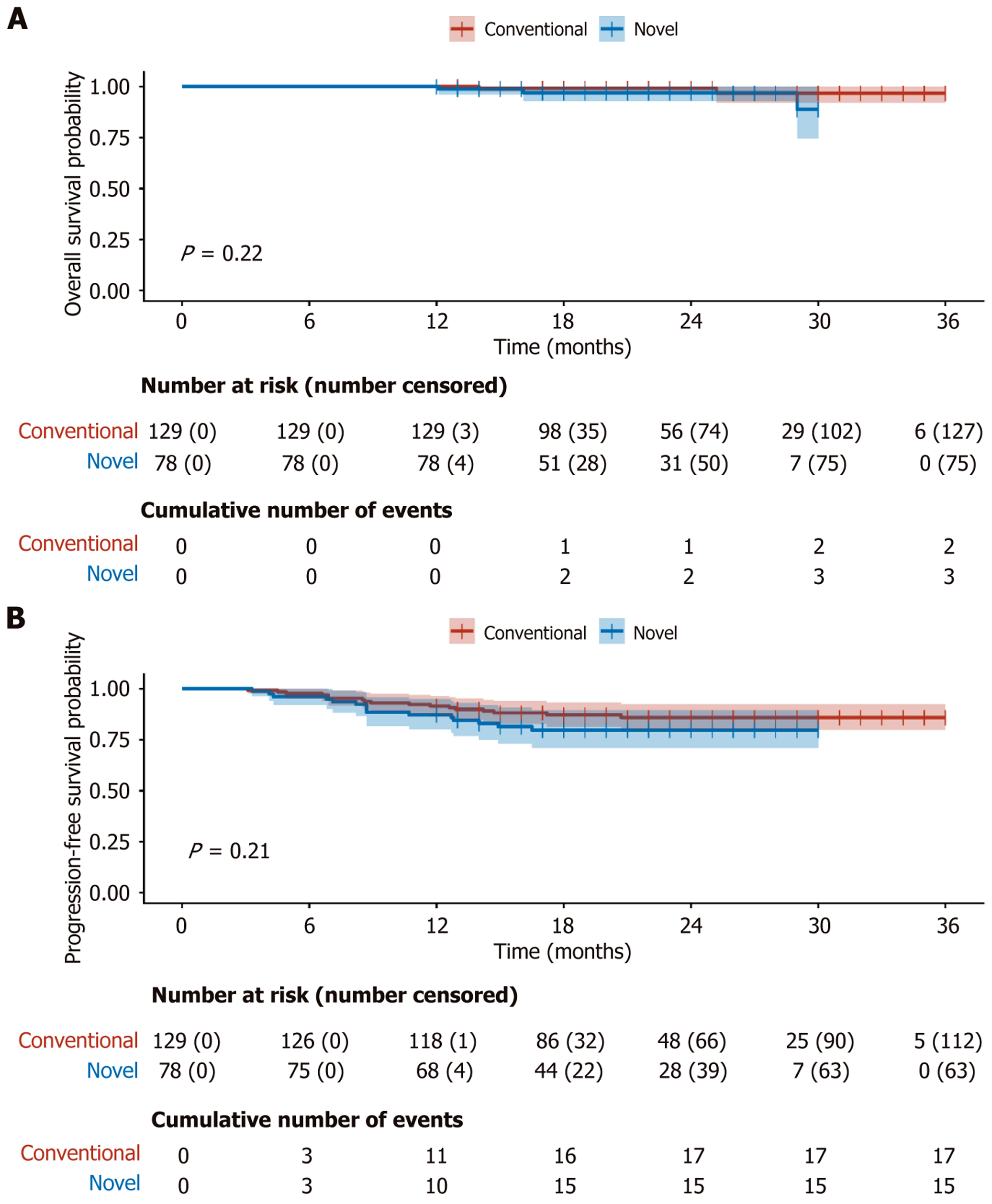
Figure 10 Exploratory Kaplan-Meier survival analysis in the comparator dataset.
A: Overall survival in the conventional and novel groups. No statistically significant difference was observed (log-rank P = 0.22); B: Progression-free survival in the conventional and novel groups. No statistically significant difference was observed (log-rank P = 0.21). Numbers at risk (number censored) are presented below each curve. Given that the comparator grouping was closely linked to anatomical subtype rather than an independent treatment strategy, these analyses should be interpreted as exploratory.
- Citation: Huang K, Hu HY, Chen K, Chen XY, Chen Z, Sun Y, Du CY, Chang GX, Fu H, Hu C, Xiao H, Lan X. Portal vein-based subsegmental classification of liver segment four for parenchyma-sparing laparoscopic resection: A retrospective study. World J Gastroenterol 2026; 32(44): 120736
- URL: https://www.wjgnet.com/1007-9327/full/v32/i44/120736.htm
- DOI: https://dx.doi.org/10.3748/wjg.120736