
















Copyright: ©Author(s) 2026.
World J Gastroenterol. Nov 28, 2026; 32(44): 120736
Published online Nov 28, 2026. doi: 10.3748/wjg.120736
Published online Nov 28, 2026. doi: 10.3748/wjg.120736
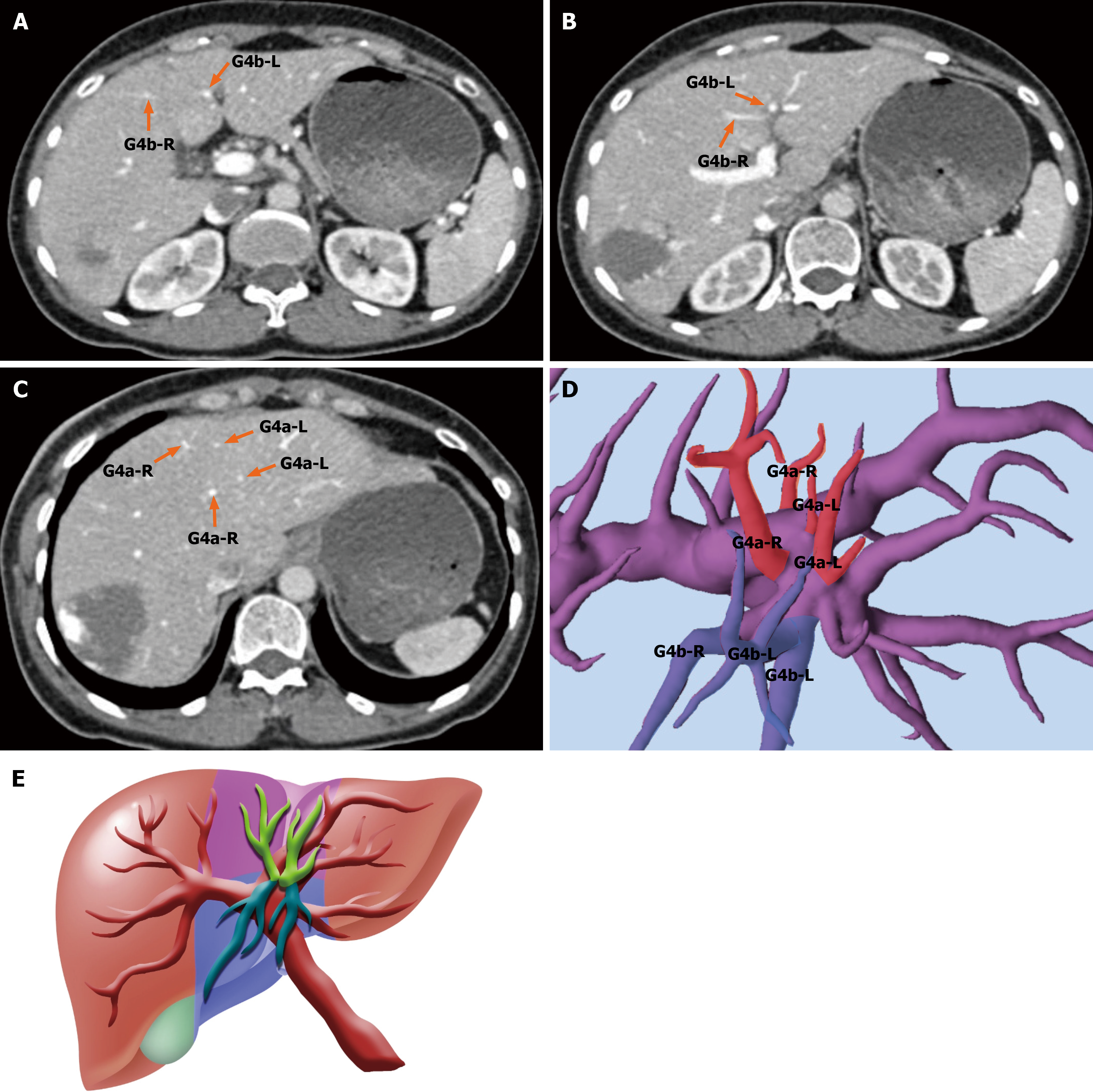
Figure 5 Type Ic classification: Fully dispersed Glissonean branching pattern in segment 4.
A-C: Axial contrast-enhanced computed tomography images from a representative type Ic case, showing fine and scattered Glissonean branches (Glissonean pedicles of segments 4a and 4b) supplying cranial and caudal subsegments without a dominant trunk; D: Three-dimensional portal vein reconstruction confirming a dispersed origin of multiple small Glissonean pedicles, each individually supplying subregions of segment 4 (S4); E: Schematic diagram illustrating the cranial-caudal perfusion pattern of S4 in type Ic, characterized by multiple independently arising Glissonean branches rather than a single dominant pedicle. G4: Glissonean pedicles of segment 4.
- Citation: Huang K, Hu HY, Chen K, Chen XY, Chen Z, Sun Y, Du CY, Chang GX, Fu H, Hu C, Xiao H, Lan X. Portal vein-based subsegmental classification of liver segment four for parenchyma-sparing laparoscopic resection: A retrospective study. World J Gastroenterol 2026; 32(44): 120736
- URL: https://www.wjgnet.com/1007-9327/full/v32/i44/120736.htm
- DOI: https://dx.doi.org/10.3748/wjg.120736