
















Copyright: ©Author(s) 2026.
World J Gastroenterol. Nov 28, 2026; 32(44): 120736
Published online Nov 28, 2026. doi: 10.3748/wjg.120736
Published online Nov 28, 2026. doi: 10.3748/wjg.120736
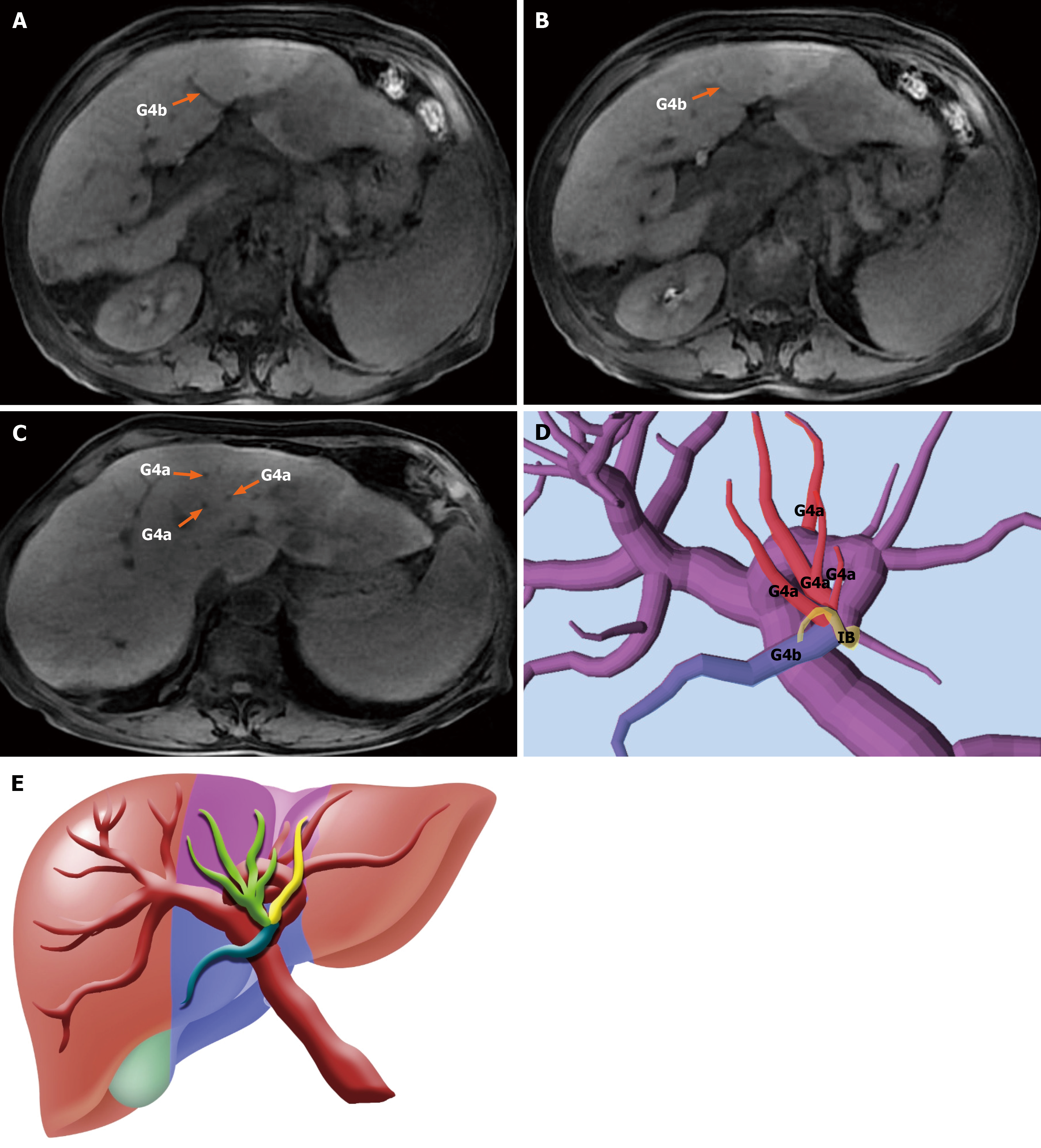
Figure 4 Type Ib classification: Unilateral cranial trunk with contralateral caudal dispersion in segment 4.
A-C: Gadolinium-enhanced magnetic resonance imaging images from a representative type Ib case. A dominant Glissonean pedicle (Glissonean pedicles of segment 4a) supplies the cranial portion of segment 4, while the caudal portion (Glissonean pedicles of segment 4b) receives scattered inflow from multiple smaller branches, indicating perfusion asymmetry; D: Three-dimensional reconstruction showing unilateral cranial inflow and contralateral caudal dispersion, with one intermediate branch arising near the bifurcation zone; E: Schematic diagram illustrating the type Ib pattern, with centralized cranial inflow and diffuse caudal distribution, reinforcing the concept of asymmetric cephalocaudal perfusion in this subtype. IB: Intermediate branch; G4: Glissonean pedicles of segment 4.
- Citation: Huang K, Hu HY, Chen K, Chen XY, Chen Z, Sun Y, Du CY, Chang GX, Fu H, Hu C, Xiao H, Lan X. Portal vein-based subsegmental classification of liver segment four for parenchyma-sparing laparoscopic resection: A retrospective study. World J Gastroenterol 2026; 32(44): 120736
- URL: https://www.wjgnet.com/1007-9327/full/v32/i44/120736.htm
- DOI: https://dx.doi.org/10.3748/wjg.120736