
















Copyright: ©Author(s) 2026.
World J Gastroenterol. Nov 14, 2026; 32(42): 117657
Published online Nov 14, 2026. doi: 10.3748/wjg.117657
Published online Nov 14, 2026. doi: 10.3748/wjg.117657
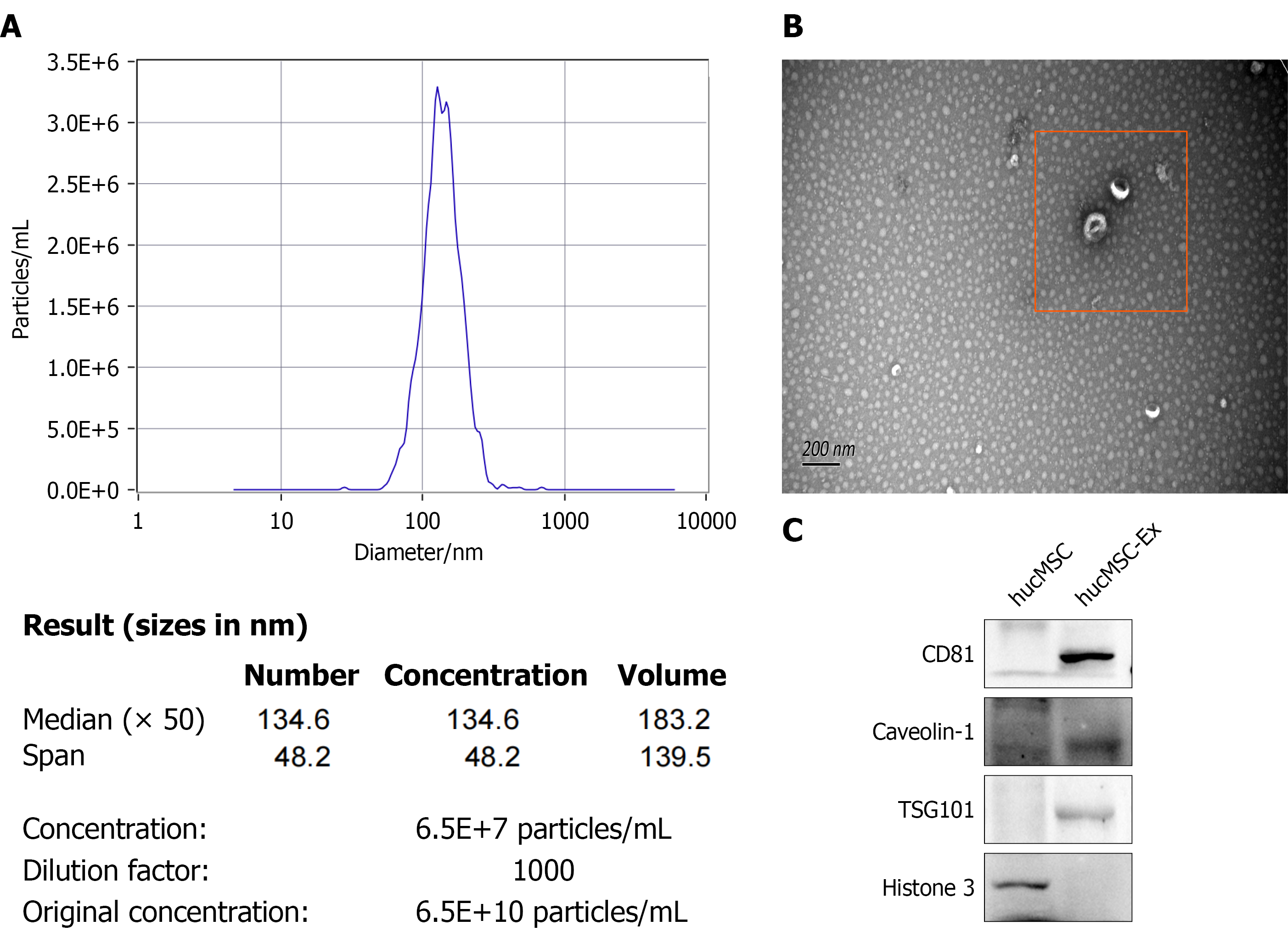
Figure 1 Identification of human umbilical cord mesenchymal stem cell-derived exosomes.
A: Size distribution and particle concentration of hucMSC-Ex determined by nanoparticle tracking analysis; B: Morphological identification of hucMSC-Ex by transmission electron microscopy (scale bar = 200 nm); C: Western blot analysis of hucMSC-Ex surface markers. CD: Cluster of differentiation; hucMSC: Human umbilical cord mesenchymal stem cell; hucMSC-Ex: Human umbilical cord mesenchymal stem cell-derived exosomes.
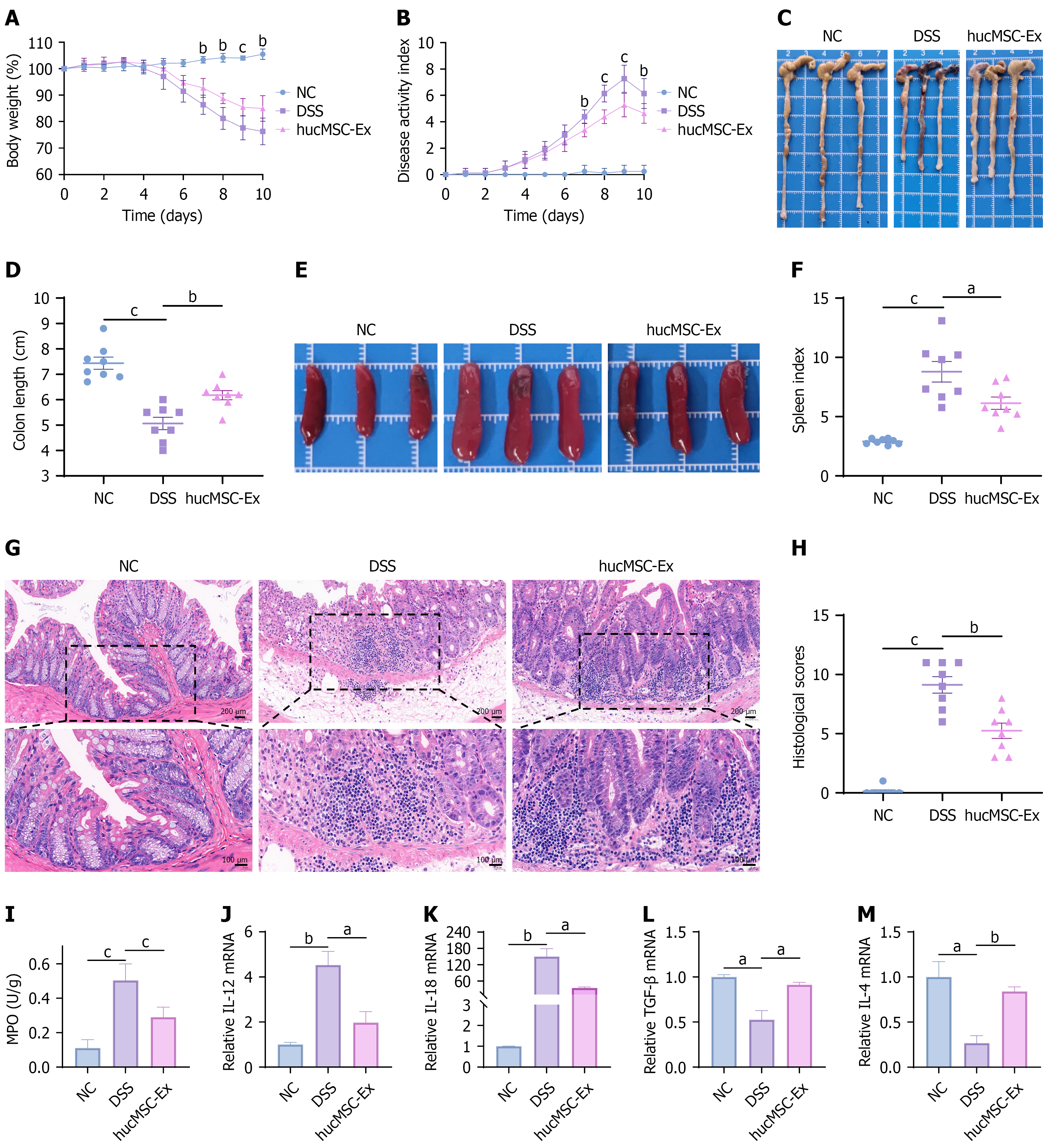
Figure 2 Human umbilical cord mesenchymal stem cell-derived exosomes attenuate dextran sulfate sodium-induced colitis in mice.
A: Daily body weight changes in each group (dextran sulfate sodium vs human umbilical cord mesenchymal stem cell-derived exosomes); B: Disease activity index (DAI) scores in each group; C: Gross appearance of representative colons from mice in each group; D: Colon lengths of mice in each group; E: Macroscopic appearance of spleens from mice in each group; F: Spleen index of mice in each group; G: Representative histopathological changes in colon tissues assessed by hematoxylin and eosin staining (20 ×, scale bar = 200 μm; 40 ×, scale bar = 100 μm); H: Histological score of colons in each group; I: Colonic myeloperoxidase activity in each group; J-M: Messenger RNA expression levels of the inflammatory cytokines IL-12, IL-18, TGF-β, and IL-4. Data are presented as the mean ± SD. aP < 0.05. bP < 0.01. cP < 0.001. dP < 0.0001. Each experiment was repeated three times independently (n = 8 mice per group). NC: Negative control; DSS: Dextran sulfate sodium; MPO: Myeloperoxidase; IL: Interleukin; mRNA: Messenger RNA; TGF: Transforming growth factor; hucMSC-Ex: Human umbilical cord mesenchymal stem cell-derived exosomes.
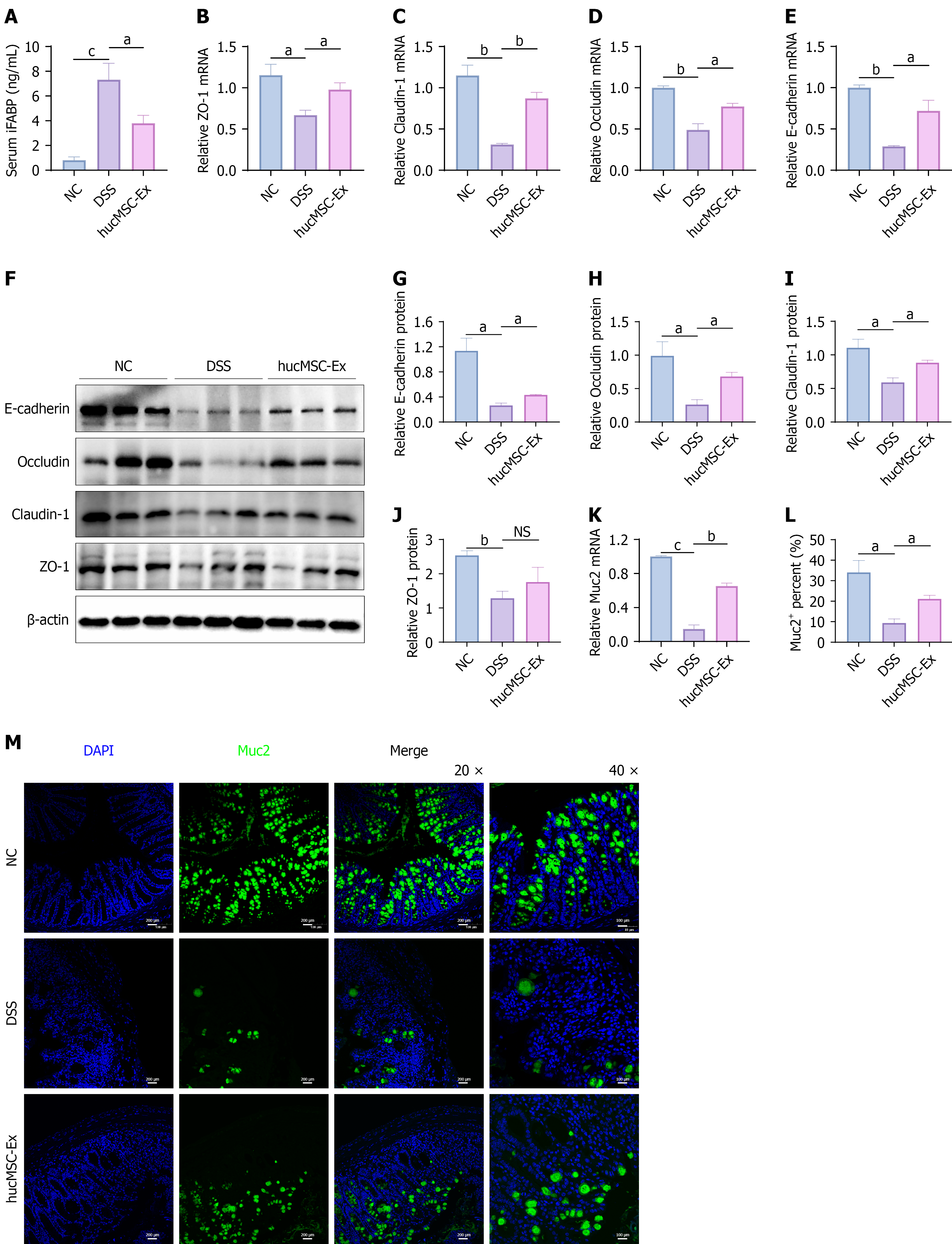
Figure 3 Human umbilical cord mesenchymal stem cell-derived exosomes alleviate intestinal mucosal barrier dysfunction in dextran sulfate sodium-induced colitis mice.
A: Serum levels of intestinal fatty acidbinding protein (iFABP) as an indicator of intestinal mucosal permeability; B-E: Messenger RNA expression levels of the tight junction proteins ZO-1, claudin-1, and occludin, and the adhesion junction protein Ecadherin; F: Representative Western blot bands showing protein expression of ZO-1, claudin-1, occludin, and E-cadherin; G-J: Quantitative analysis of protein expression for ZO-1, occludin, claudin-1, and E-cadherin by Western blot; K: Messenger RNA expression level of mucin 2 (Muc2) (20 ×, scale bar = 200 μm; 40 ×, scale bar = 100 μm); L and M: Immunofluorescence staining for Muc2 in colonic sections. Data are presented as the mean ± SD. aP < 0.05. bP < 0.01. cP < 0.001. dP < 0.0001. Each experiment was repeated three times independently (n = 8 mice per group). NC: Negative control; DSS: Dextran sulfate sodium; hucMSC-Ex: Human umbilical cord mesenchymal stem cell-derived exosomes; NS: No significance; mRNA: Messenger RNA; ZO-1: Zonula occludens-1; iFABP: Intestinal fatty acid-binding protein; DAPI: 4’,6-diamidino-2-phenylindole; Muc2: Mucin 2.
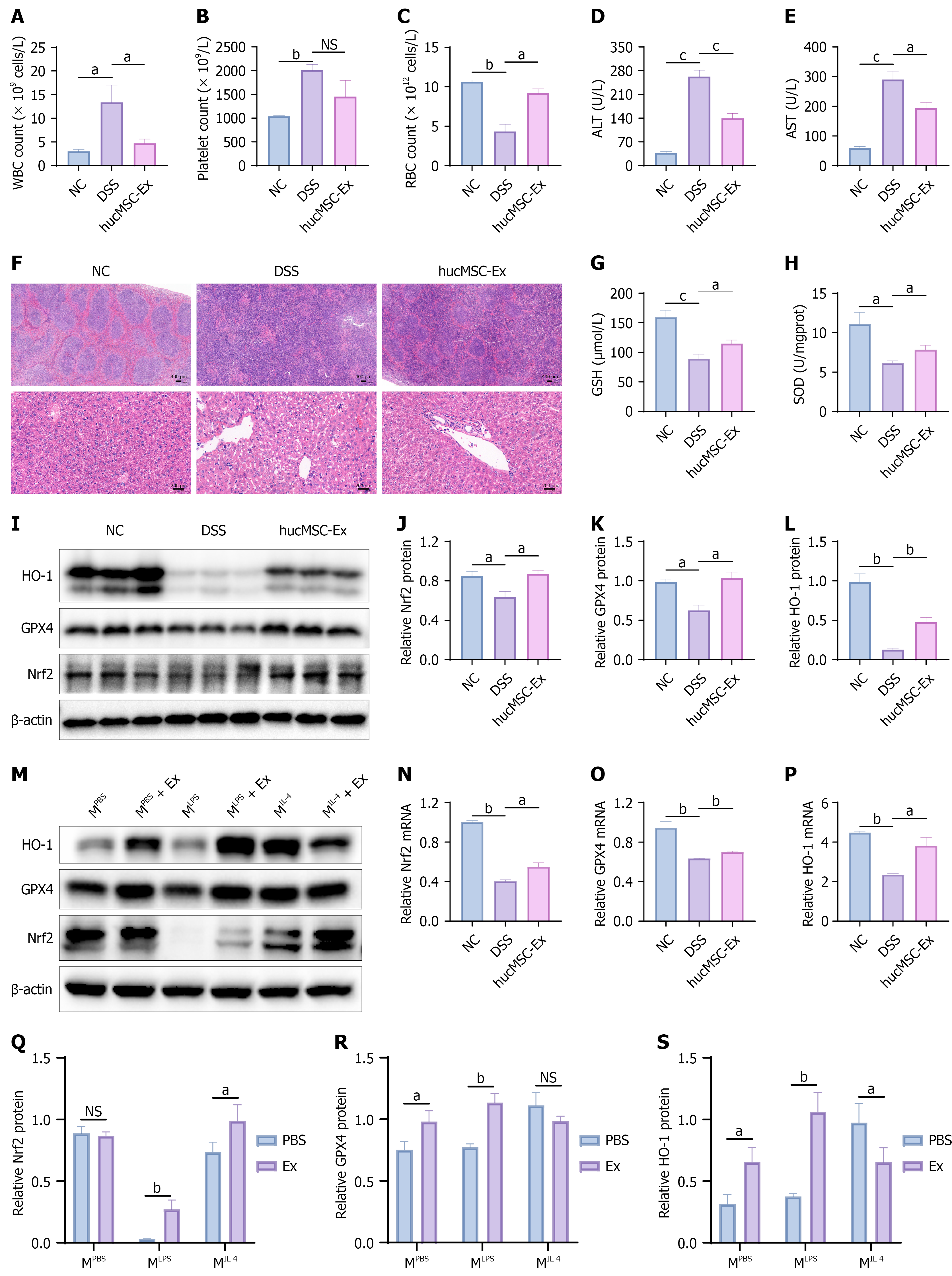
Figure 4 Human umbilical cord mesenchymal stem cell-derived exosomes upregulate the nuclear factor-erythroid 2 related factor 2/heme oxygenase-1/glutathione peroxidase 4 axis to reduce inflammation and oxidative stress.
A-C: Routine whole blood parameters as indicators of systemic inflammation; D and E: Serum alanine aminotransferase and aspartate aminotransferase levels as markers of liver function; F: Hematoxylin-eosin staining of mouse spleen (5 ×, scale bar = 400 μm) and liver tissues (30 ×, scale bar = 200 μm); G and H: Activities of the antioxidant markers glutathione and superoxide dismutase in colon tissues; I: Protein expression levels of the nuclear factor-erythroid 2 related factor 2 (Nrf2)/heme oxygenase-1 (HO-1)/glutathione peroxidase 4 (GPX4) axis in mouse colon tissues; J-L: Quantitative analysis of protein expression of Nrf2, HO-1, and GPX4 by Western blot; M: Western blot analysis of Nrf2, HO-1, and GPX4 expression in RAW264.7 cells after human umbilical cord mesenchymal stem cell-derived exosome (hucMSC-Ex) treatment; N-P: Messenger RNA expression levels of Nrf2, HO-1, and GPX4 in mouse colon tissues; Q-S: Quantitative analysis of protein expression of Nrf2, HO-1, and GPX4 in RAW264.7 cells treated with hucMSC-Ex. aP < 0.05. bP < 0.01. cP < 0.001. NS: No significance; NC: Negative control; DSS: Dextran sulfate sodium; hucMSC-Ex: Human umbilical cord mesenchymal stem cell-derived exosomes; WBC: White blood cell; RBC: Red blood cell; ALT: Alanine aminotransferase; AST: Aspartate aminotransferase; GSH: Glutathione; SOD: Superoxide dismutase; Nrf2: Nuclear factor-erythroid 2 related factor 2; HO-1: Heme oxygenase-1; GPX4: Glutathione peroxidase 4; MPBS: Phosphate-buffered saline treatment; MLPS: Lipopolysaccharide + interferon-γ treatment; MIL-4: Interleukin-4 + interleukin-13 treatment; Ex: Exosomes; PBS: Phosphate-buffered saline.
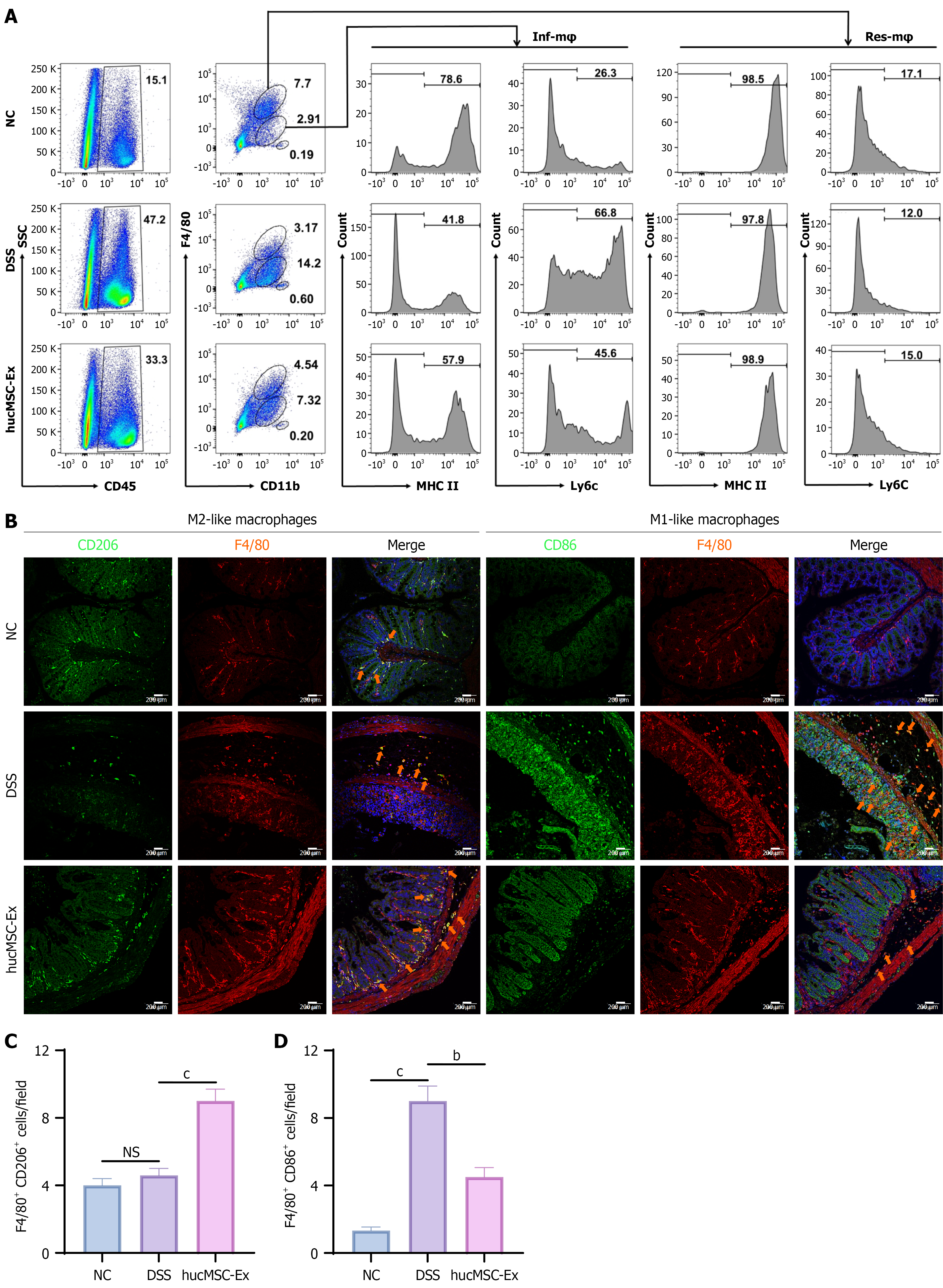
Figure 5 Human umbilical cord mesenchymal stem cell-derived exosomes modulate the plasticity of intestinal macrophages.
A: Flow cytometric analysis of intestinal macrophages in colon tissues stained with anti-F4/80, anti-CD11b, anti-MHCII, and anti-Ly6C antibodies; B: Immunofluorescence (IF) staining of pro-resolving macrophages (F4/80+ CD206+) and pro-inflammatory macrophages (F4/80+ CD86+) by immunofluorescence (IF) in colon tissues (20 ×, scale bar = 200 μm); C: Quantification of F4/80+ CD206+ cells in colon tissues; D: Quantification of F4/80+ CD86+ cells in colon tissues. Data are presented as the mean ± SD. bP < 0.01. cP < 0.001. Each experiment was repeated three times independently (n = 8 mice per group). NS: No significance; NC: Negative control; DSS: Dextran sulfate sodium; hucMSC-Ex: Human umbilical cord mesenchymal stem cell-derived exosomes; CD: Cluster of differentiation; MHC: Major histocompatibility complex; Ly6C: Lymphocyte antigen 6C; Inf-Mφ: Inflammatory macrophages; Res-Mφ: Resident macrophages.
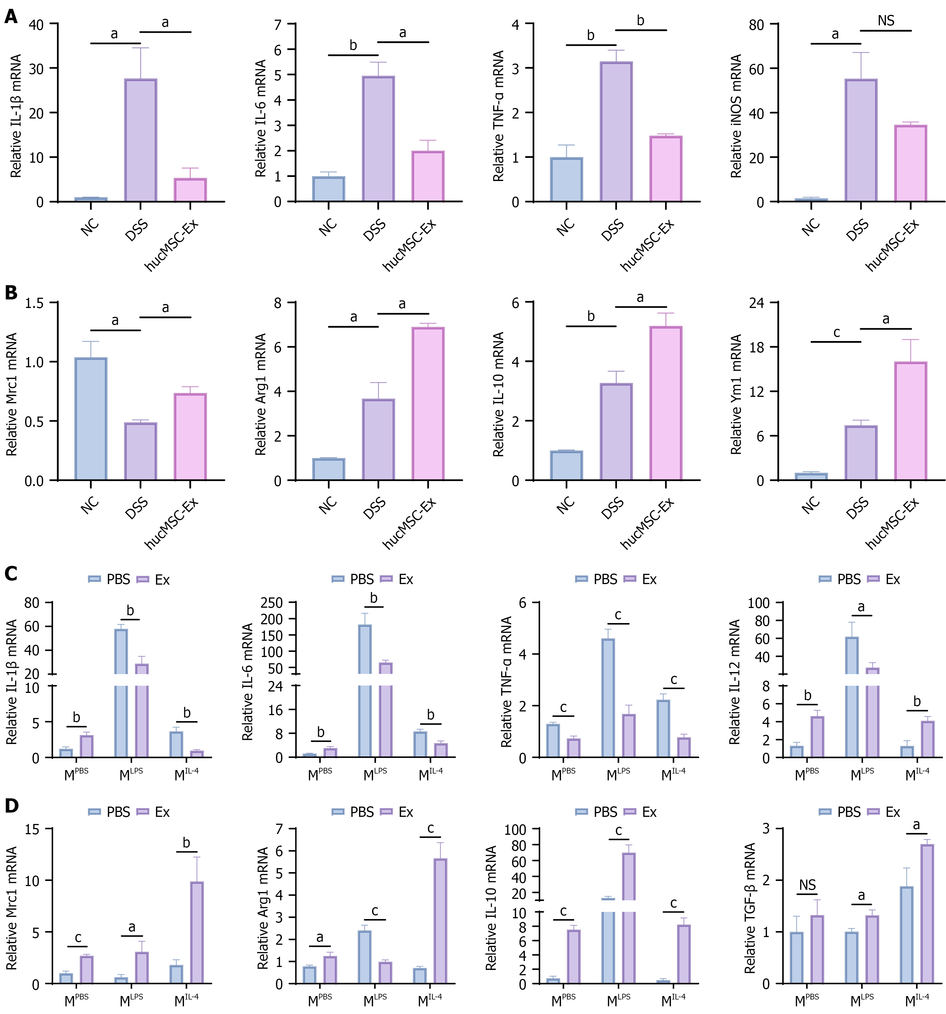
Figure 6 Human umbilical cord mesenchymal stem cell-derived exosomes promote transformation of macrophage subsets.
A and B: Relative mRNA expression levels of the inflammatory cytokines interleukin (IL)-1β, IL-6, tumor necrosis factor (TNF)-α, inducible nitric oxide synthase, Mrc1, Arg1, IL-10, and Ym1 determined by reverse transcription quantitative polymerase chain reaction (RT-qPCR); C and D: Relative mRNA expression of the M1-related genes IL-1β, IL-6, TNF-α, and IL-12, and the M2-related genes Mrc1, Arg1, IL-10, and transforming growth factor-β determined by RT-qPCR in RAW264.7 cells stimulated with phosphate-buffered saline, lipopolysaccharide + interferon-γ, and IL-4 + IL-13 following human umbilical cord mesenchymal stem cell-derived exosome treatment. Data are presented as the mean ± SD. Each experiment was repeated three times independently (n = 8 mice per group). aP < 0.05. bP < 0.01. cP < 0.001. NS: No significance; NC: Negative control; DSS: Dextran sulfate sodium; hucMSC-Ex: Human umbilical cord mesenchymal stem cell-derived exosomes; IL: Interleukin; mRNA: Messenger RNA; TNF: Tumor necrosis factor; iNOS: Inducible nitric oxide synthase; TGF: Transforming growth factor; MPBS: Phosphate-buffered saline treatment; MLPS: Lipopolysaccharide + interferon-γ treatment; MIL-4: Interleukin-4 + interleukin-13 treatment; Ex: Exosomes; PBS: Phosphate-buffered saline.
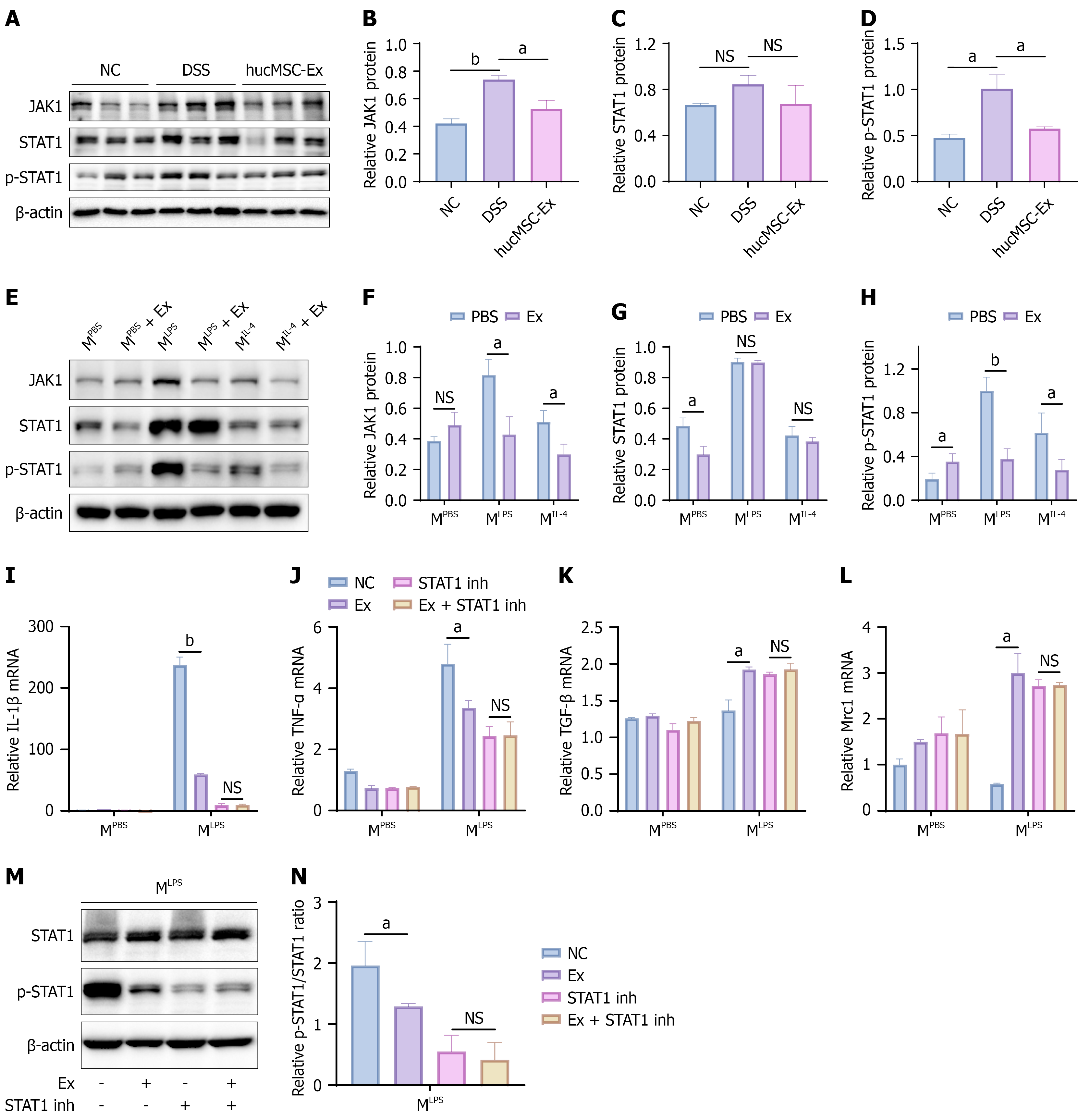
Figure 7 Human umbilical cord mesenchymal stem cell-derived exosomes regulate macrophage phenotype via the Janus kinase 1/ signal transducer and activator of transcription 1 signaling pathway.
A-D: Western blot analysis of Janus kinase (JAK) 1, signal transducer and activator of transcription (STAT) 1, and phospho-STAT1 (p-STAT1) protein expression in colon tissues; E-H: Western blot analysis of JAK1, STAT1, and p-STAT1 protein expression in RAW264.7 cells treated with human umbilical cord mesenchymal stem cell-derived exosomes (hucMSC-Ex); I-L: Determination of interleukin-1β, tumor necrosis factor-α, transforming growth factor-β, and Mrc1 mRNA expression by reverse transcription quantitative polymerase chain reaction after STAT1 inhibition followed by hucMSC-Ex administration; M and N: Determination of protein expression of p-STAT1 and STAT1 by Western blot after STAT1 inhibition followed by hucMSC-Ex administration. Data are presented as the mean ± SD. Each experiment was repeated three times independently (n = 8 mice per group). aP < 0.05. bP < 0.01. NS: No significance; NC: Negative control; DSS: Dextran sulfate sodium; hucMSC-Ex: Human umbilical cord mesenchymal stem cell-derived exosomes; JAK: Janus kinase; STAT: Signal transducer and activator of transcription; p-STAT: Phospho-signal transducer and activator of transcription; MPBS: Phosphate-buffered saline treatment; MLPS: Lipopolysaccharide + interferon-γ treatment; MIL-4: Interleukin-4 + interleukin-13 treatment; Ex: Exosomes; PBS: Phosphate-buffered saline; IL: Interleukin; mRNA: Messenger RNA; TNF: Tumor necrosis factor; TGF: Transforming growth factor; STAT1 inh: Signal transducer and activator of transcription 1 inhibitor.
- Citation: Tian LL, Zhu J, Fan ZZ, Shi YT, Zhao LQ, He JD, Liang J. Human umbilical cord mesenchymal stem cell-derived exosomes alleviate experimental colitis by maintaining the intestinal barrier and remodeling macrophage polarization. World J Gastroenterol 2026; 32(42): 117657
- URL: https://www.wjgnet.com/1007-9327/full/v32/i42/117657.htm
- DOI: https://dx.doi.org/10.3748/wjg.117657