
















Copyright: ©Author(s) 2026.
World J Gastroenterol. Sep 21, 2026; 32(35): 121494
Published online Sep 21, 2026. doi: 10.3748/wjg.121494
Published online Sep 21, 2026. doi: 10.3748/wjg.121494
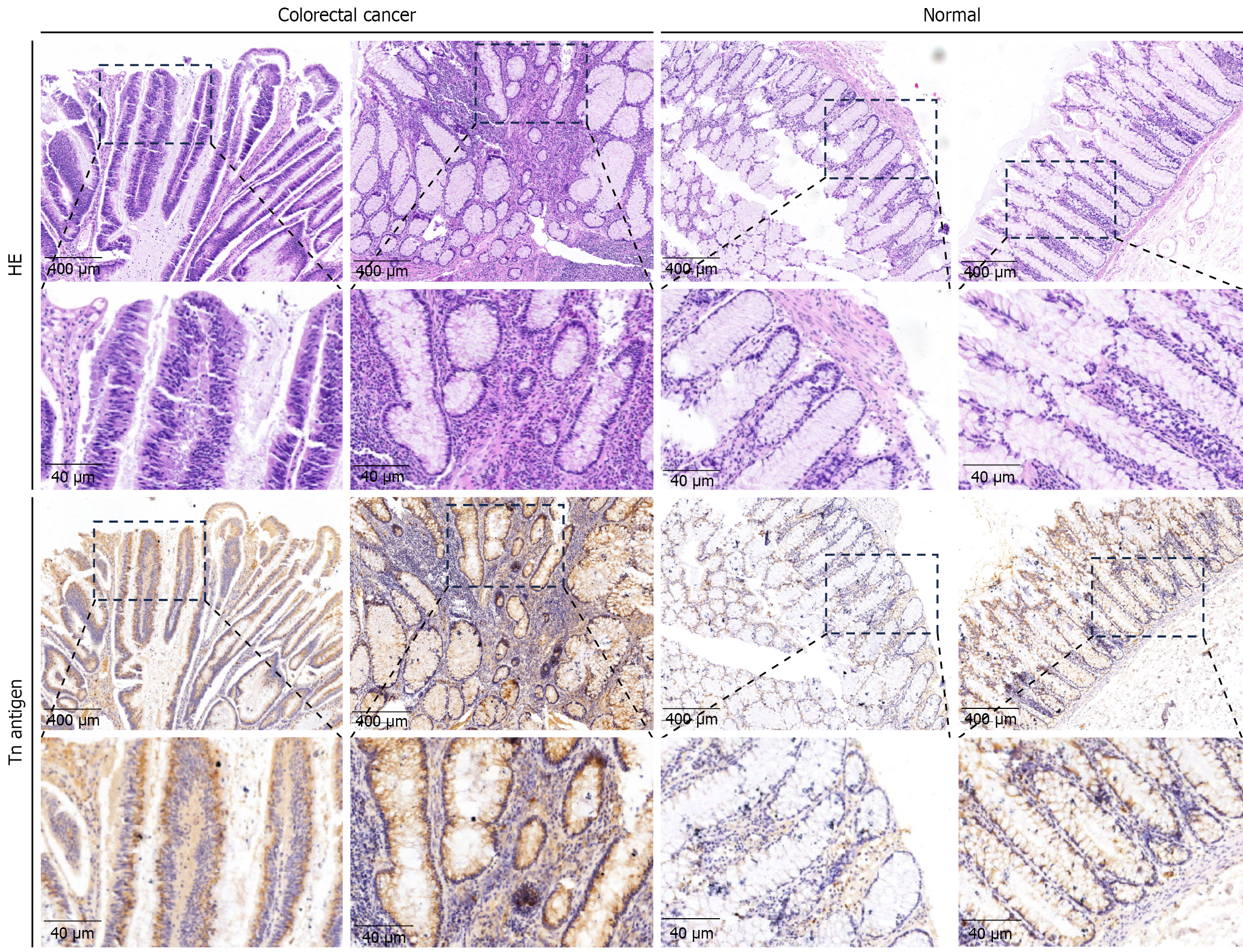
Figure 1 Immunohistochemical detection of Tn antigen expression in colorectal cancer tissues.
HE: Hematoxylin-eosin.
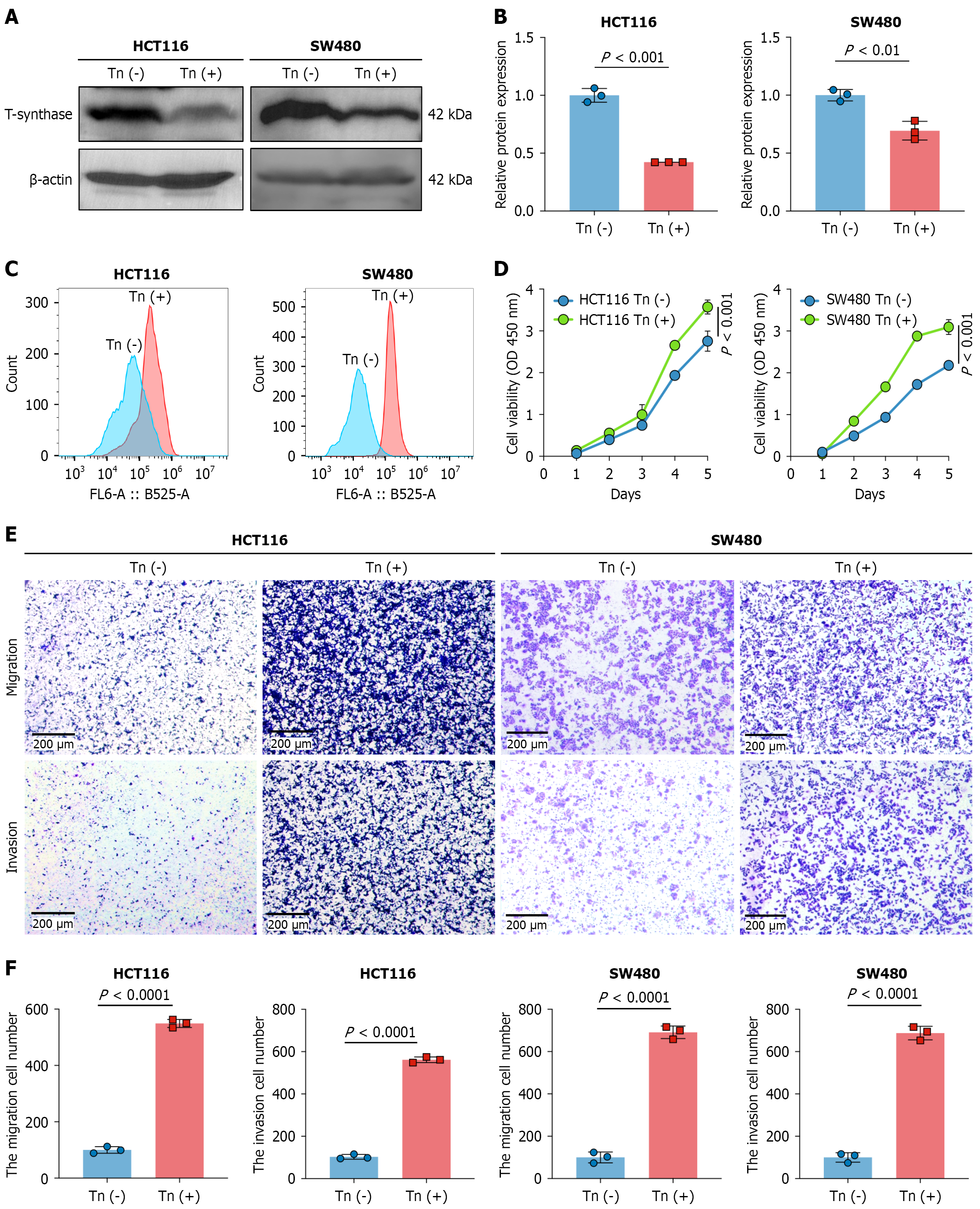
Figure 2 High expression of the Tn antigen increases the proliferation, invasion, and migration capabilities of colorectal cancer cells.
A and B: Western blot analysis of Tn antigen expression; C: Flow cytometry detection of Tn antigen expression; D: Analysis by a cell counting kit-8 proliferation assay revealed a significant increase in the proliferation rate of T-synthase-knockout cells; E and F: Analysis by Transwell assay indicates increased invasion ability of Tn (+) colorectal cancer cells. OD: Optical density.
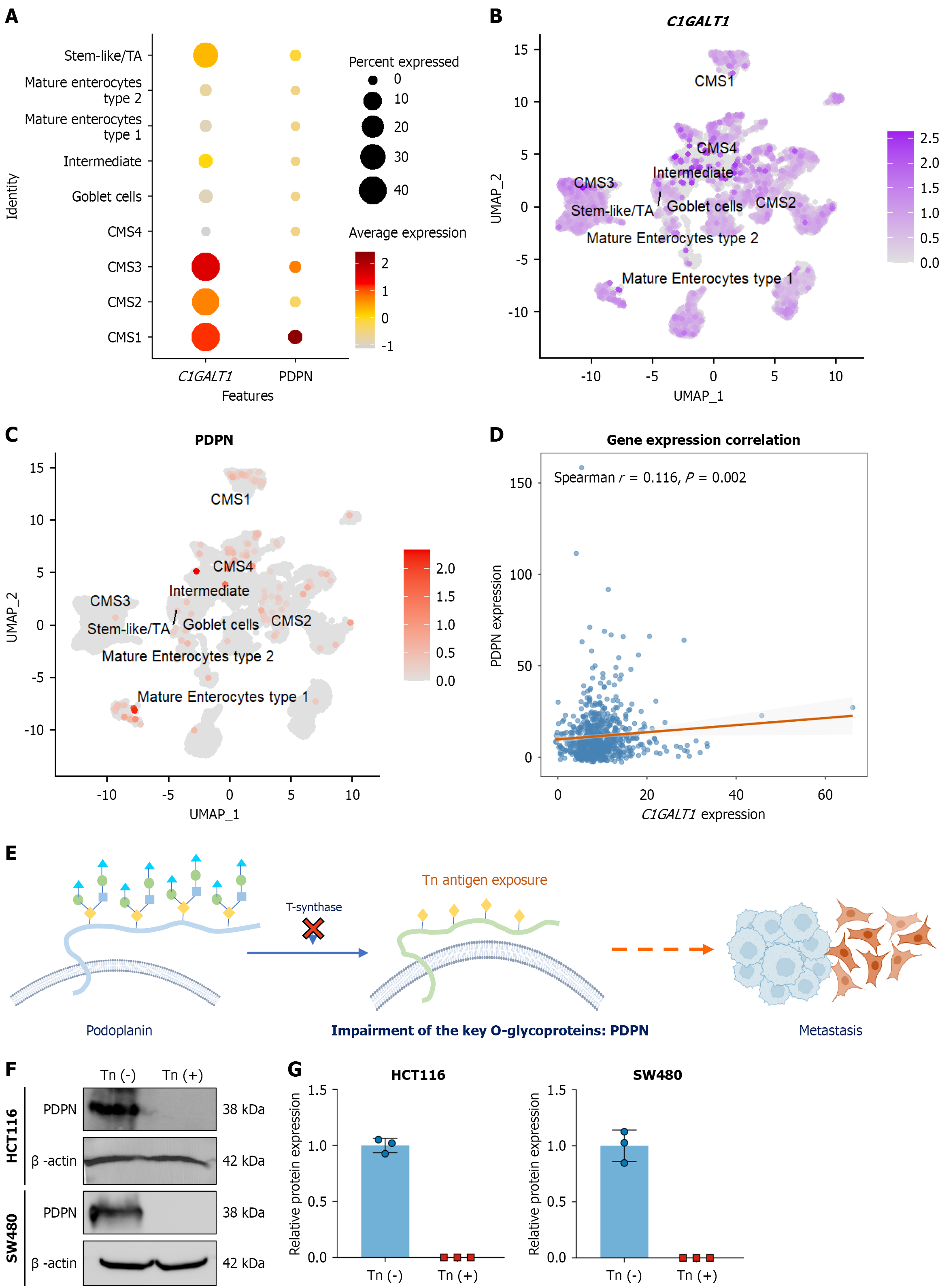
Figure 3 Relationships between Tn antigen and podoplanin expression.
A: Expression characteristics of C1GALT1 and podoplanin (PDPN) in major cellular subsets; B and C: Spatial expression localization of C1GALT1 and PDPN in the single-cell landscape; D: Scatter plot of C1GALT1 vs PDPN expression; E: Mechanistic role of PDPN; F and G: Western blot analysis confirming reduced PDPN expression in Tn antigen-expressing. CMS: Consensus molecular subtype; PDPN: Podoplanin.
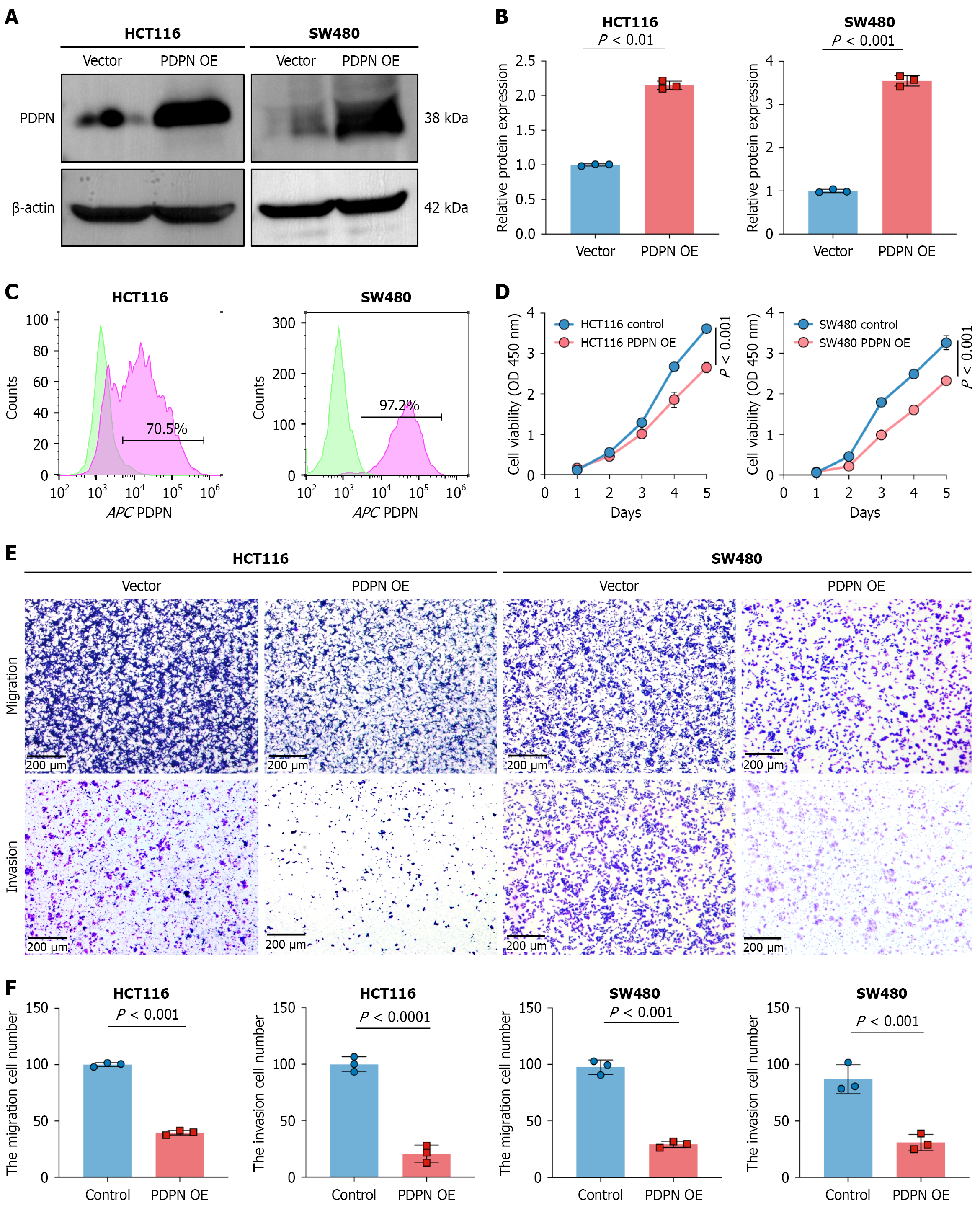
Figure 4 Overexpression of podoplanin decreases the proliferation and metastatic capabilities of colorectal cancer cells.
A and B: Western blot detection of podoplanin (PDPN) overexpression; C: Flow cytometry detection of PDPN overexpression; D: The cell counting kit-8 proliferation assay revealed a significant decrease in the proliferation rate of colorectal cancer (CRC) cells overexpressing PDPN; E and F: The Transwell assay revealed a reduction in the invasion ability of CRC cells overexpressing PDPN. OE: Overexpression; PDPN: Podoplanin.
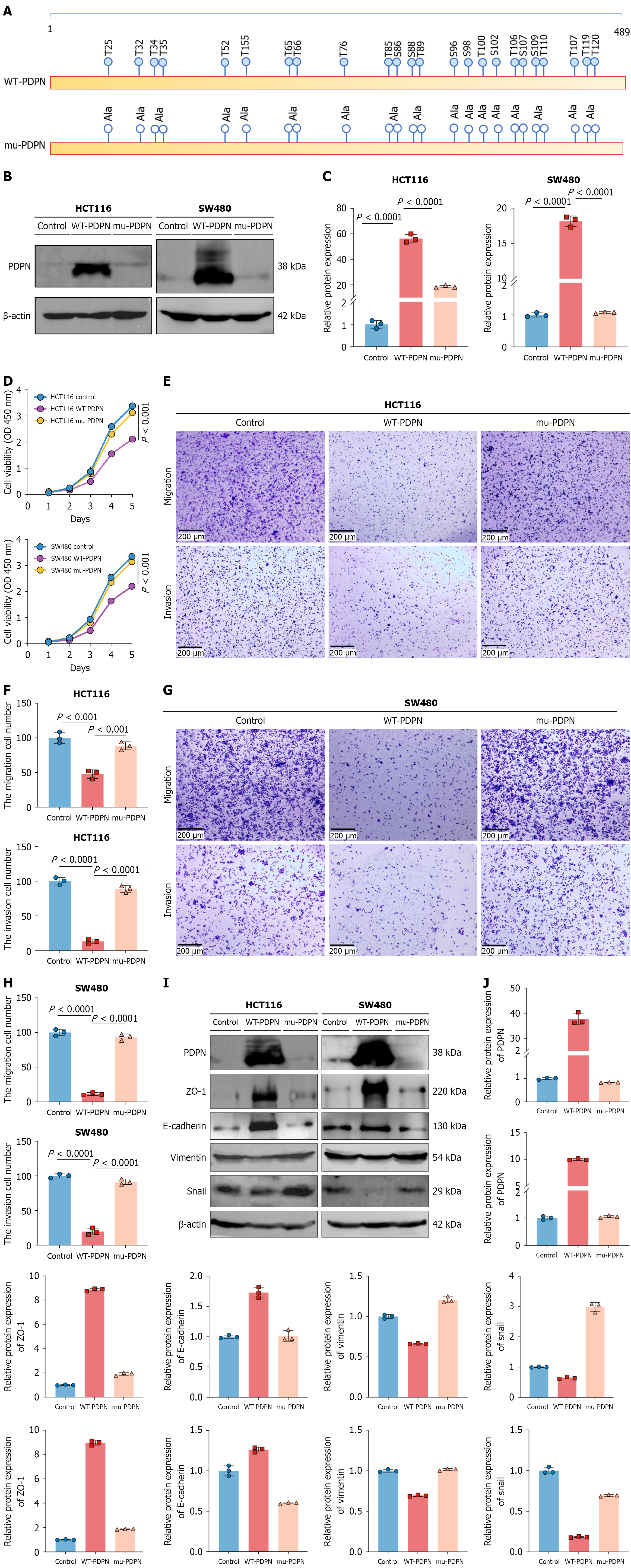
Figure 5 O-glycosylation is crucial for the function of podoplanin.
A: Schematic diagram of O-glycosylation sites on podoplanin (PDPN); B and C: Western blot validation of the expression of control, wild-type (WT), and O-glycosylation-mutated (mu) PDPN; D: The cell counting kit-8 proliferation assay; E-H: Transwell assay analysis of the migration and invasion capabilities of colorectal cancer cells overexpressing PDPN; I and J: Western blot results indicate that WT-PDPN-expressing cells can inhibit the epithelial-mesenchymal transition (EMT) process, while mu-PDPN-expressing cells promote the EMT process similarly to the control cells. Ala: Alanine; PDPN: Podoplanin; WT: Wild-type; mu: O-glycosylation-mutated; ZO-1: Zonula occludens-1.
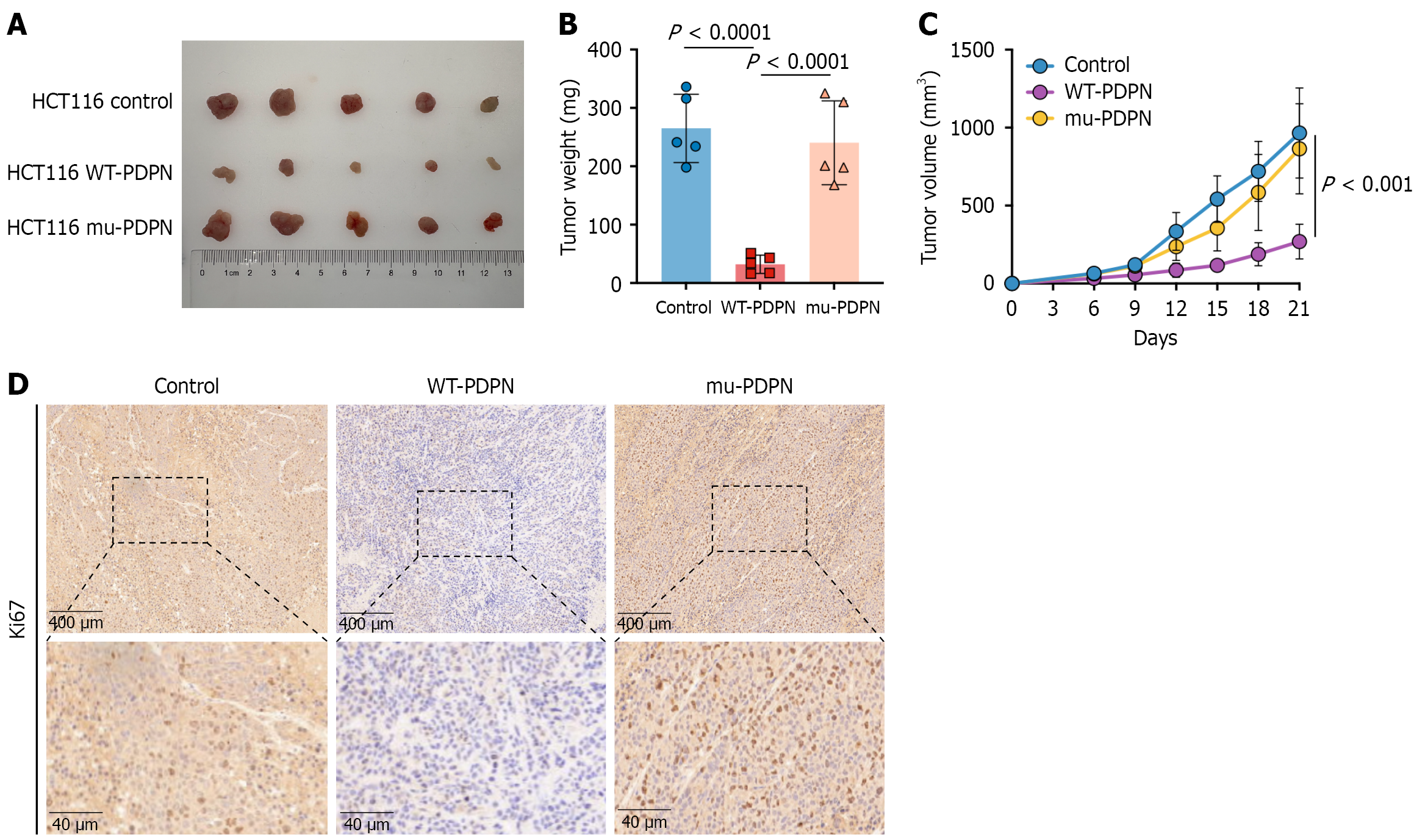
Figure 6 O-glycosylation-deficient podoplanin promotes tumor growth.
A: Effect of abnormal O-glycosylation of podoplanin on tumor growth and proliferation in a BALB/c nude mouse model; B: Tumor volume comparison; C: Tumor growth curve; D: Cell proliferation in tumors was assessed by immuno
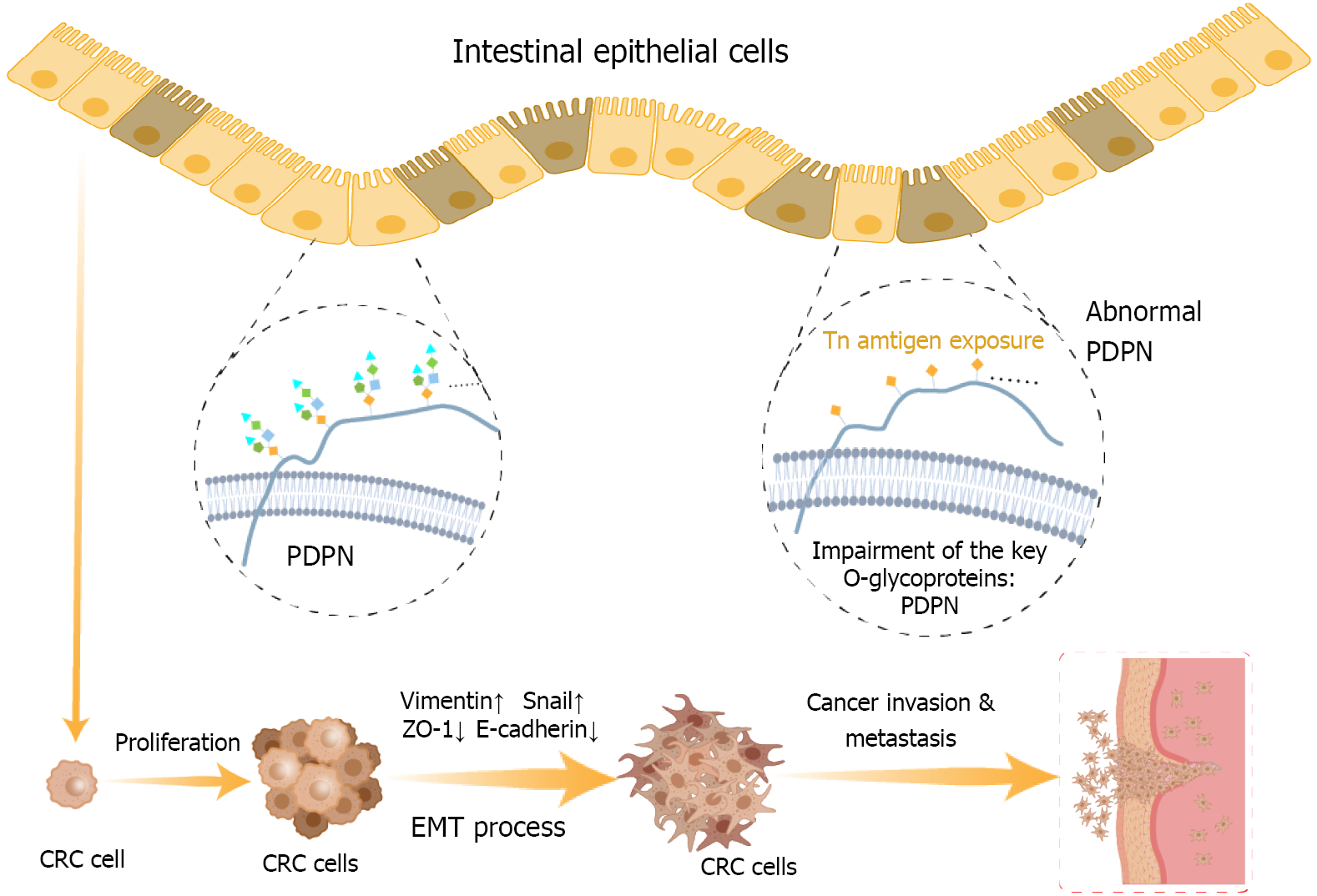
Figure 7 Aberrant O-glycosylation characterized by high Tn antigen expression drives colorectal cancer progression through the downregulation of podoplanin expression.
PDPN: Podoplanin; CRC: Colorectal cancer; EMT: Epithelial-mesenchymal transition; ZO-1: Zonula occludens-1.
- Citation: Fan LW, Wu XZ, Liu ZW, Lin Q, Du JX, Tian YT, Dong XC, Xu DK. Tn antigen-driven downregulation of podoplanin promotes colorectal cancer invasion. World J Gastroenterol 2026; 32(35): 121494
- URL: https://www.wjgnet.com/1007-9327/full/v32/i35/121494.htm
- DOI: https://dx.doi.org/10.3748/wjg.121494