
















Copyright: ©Author(s) 2026.
World J Gastroenterol. Aug 21, 2026; 32(31): 117409
Published online Aug 21, 2026. doi: 10.3748/wjg.117409
Published online Aug 21, 2026. doi: 10.3748/wjg.117409
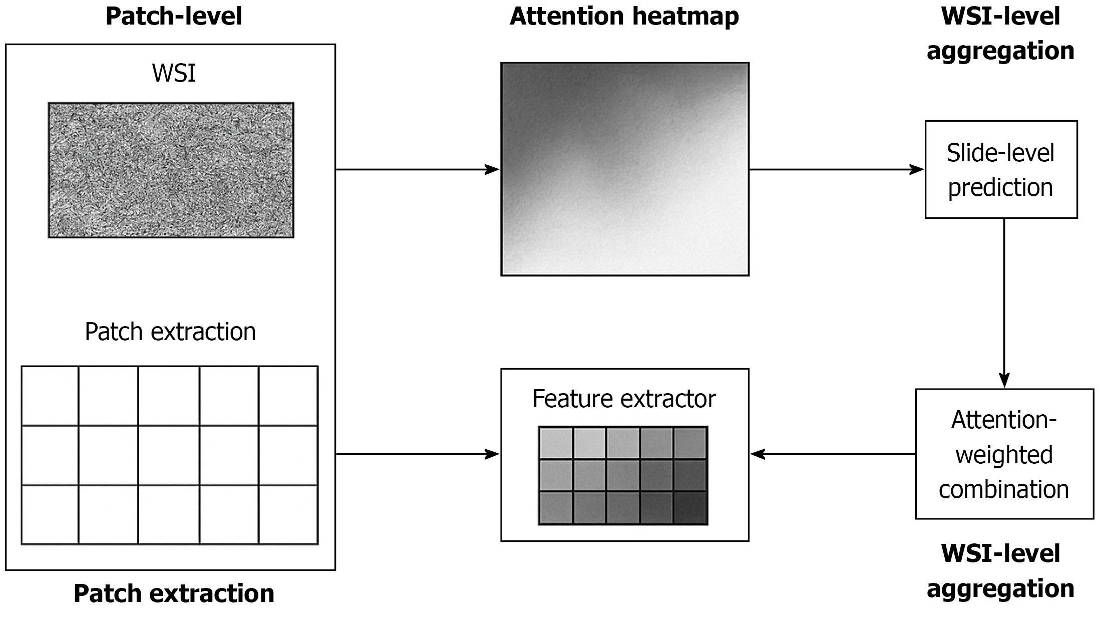
Figure 1 Patch-level feature extraction, attention-weighted aggregation, and slide-level prediction in a multiple instance learning pipeline for whole-slide pathology.
Whole-slide images are partitioned into patches, which are encoded into feature embeddings by a convolutional or transformer-based extractor. An attention module assigns weights reflecting each patch’s morphological relevance to metastatic biology, producing an interpretable attention heatmap. Weighted patch embeddings are aggregated into a slide-level representation supporting final lymph node metastasis prediction. This mechanism illustrates how multiple instance learning preserves spatial heterogeneity, highlights diagnostically salient regions, and enhances interpretability. The workflow reflects the methodological strategy implemented by Zou et al[6], forming the morphologic pillar upon which multimodal fusion with clinical and host systemic variables can be constructed. WSI: Whole-slide images.
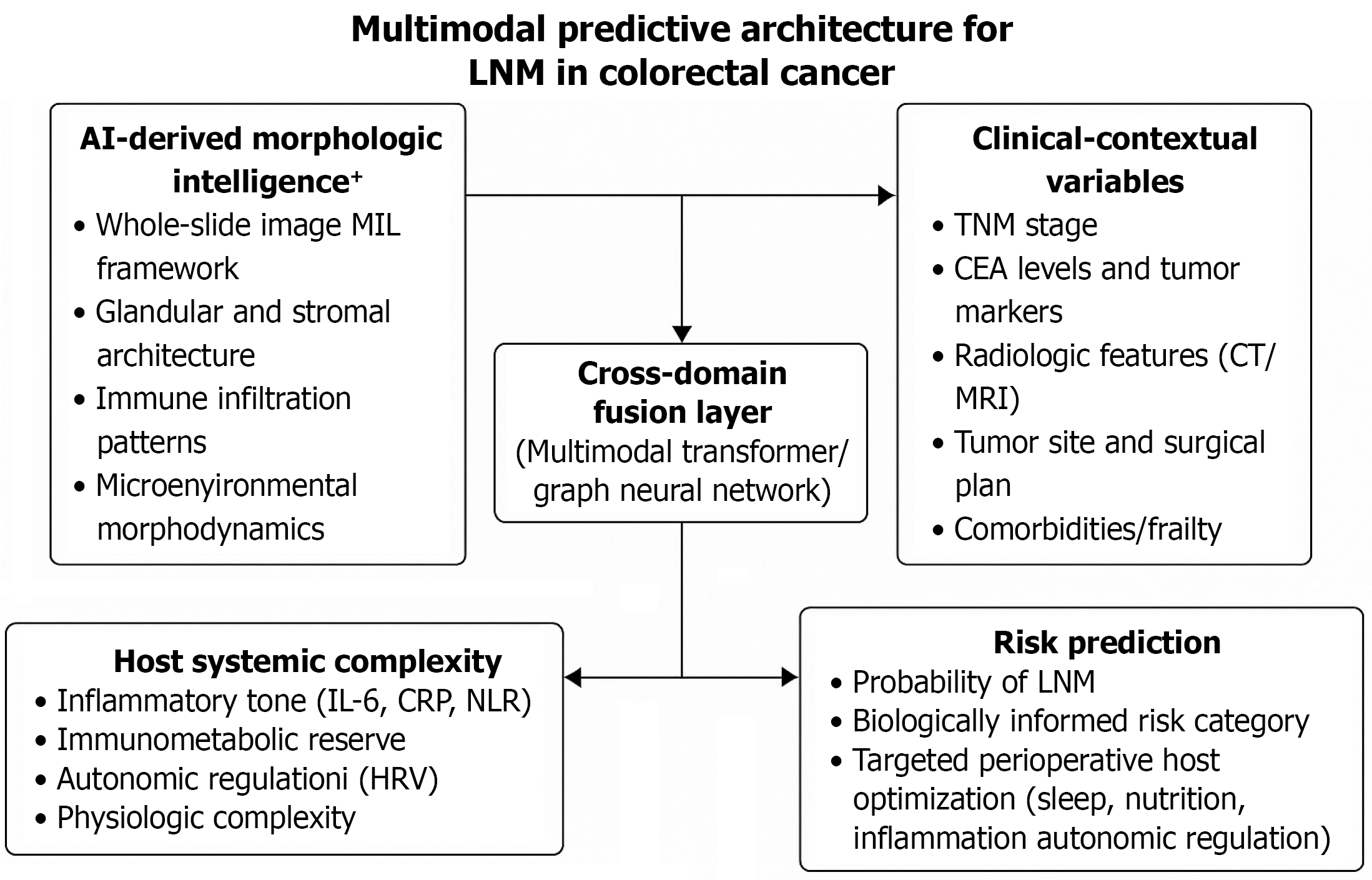
Figure 2 Multimodal predictive architecture for lymph node metastasis in colorectal cancer.
Multimodal predictive architecture extending the multiple instance learning framework reported by Zou et al[6] for lymph node metastasis in colorectal cancer. This conceptual framework builds upon AI-derived morphologic intelligence from whole-slide images, as implemented in the MIL model of Zou et al[6], and integrates two additional domains: (1) Clinical-contextual variables, including tumor-node-metastasis stage, tumor markers (carcinoembryonic antigen), radiologic features (computed tomography, magnetic resonance imaging), and comorbidities; and (2) Host systemic complexity, such as inflammatory tone (interleukin-6, C-reactive protein), heart rate variability, and immune response (neutrophil-lymphocyte ratio). A cross-domain fusion layer extends the morphology-based prediction paradigm toward a biologically integrated model of lymph node metastasis risk, supporting more comprehensive and individualized surgical decision-making. AI: Artificial intelligence; WSI: Whole-slide images; IL: Interleukin; CRP: C-reactive protein; HRV: Heart rate variability; NLR: Neutrophil-lymphocyte ratio; TNM: Tumor-node-metastasis; CEA: Carcinoembryonic antigen; CT: Computed tomography; MRI: Magnetic resonance imaging; LNM: Lymph node metastasis.
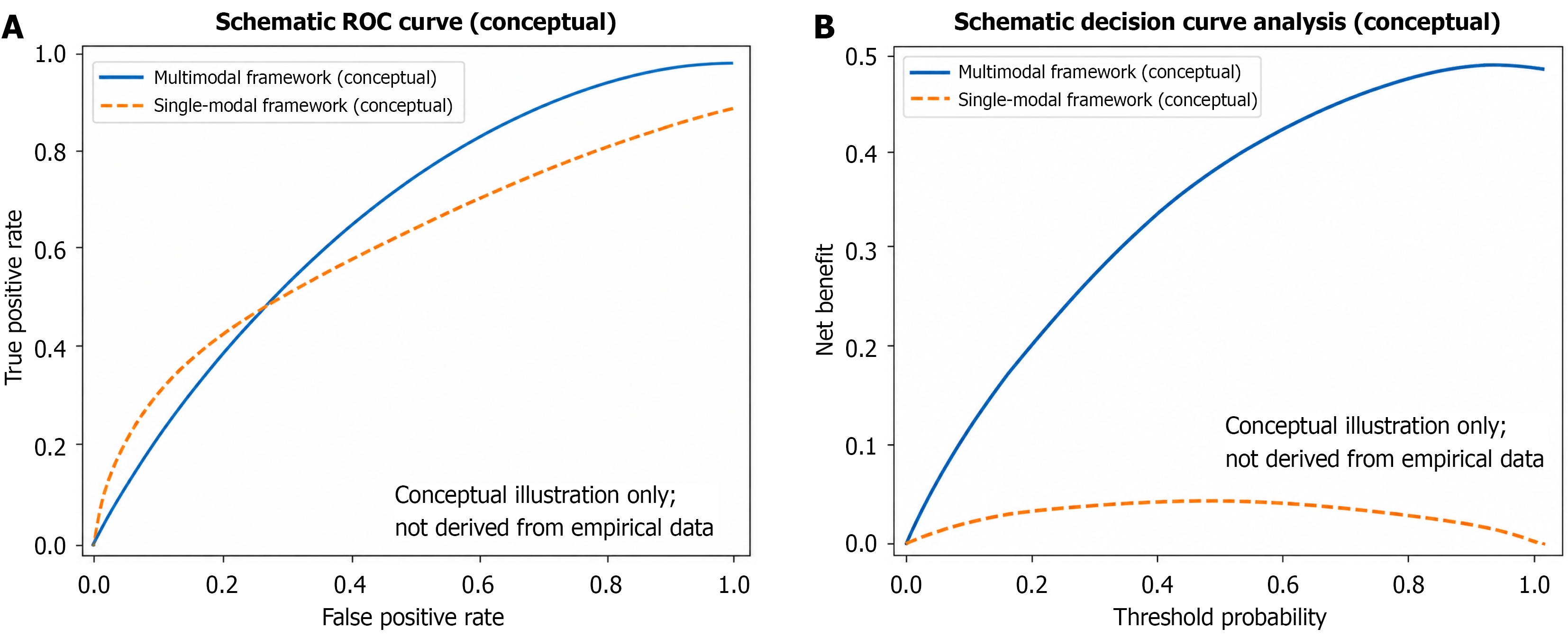
Figure 3 Conceptual comparison of single-modal vs multimodal prediction frameworks: Schematic receiver operating characteristic discrimination and decision-curve utility.
A: Schematic receiver operating characteristic curves illustrate the conceptual gain in discrimination when artificial intelligence pathology is hypothetically combined with clinical and host systemic variables; B: Schematic decision curve analysis illustrates the conceptual increase in net benefit across a range of threshold probabilities for hypothetical clinical decision scenarios. All curves are purely illustrative and are not derived from any empirical dataset; they do not report observed area under the curve, calibration, net benefit, or any other performance metric. Accordingly, the axes and curve shapes are intended only to convey qualitative relationships and should not be interpreted as dataset-specific evidence or quantitative support for any clinical action. The morphology-only curve is shown for conceptual reference to the multiple instance learning-based approach discussed by Zou et al[6]; the multimodal curves illustrate the proposed extended capabilities of an integrated predictive framework. Examples of potential decision contexts (e.g., surgical planning, neoadjuvant strategy, surveillance intensity) are provided for illustration only and do not imply validated thresholds or prescriptive recommendations. ROC: Receiver operating characteristic.
- Citation: Wang G, Pan SJ. Artificial intelligence morphology and host complexity for precision prediction of nodal metastasis in colorectal cancer. World J Gastroenterol 2026; 32(31): 117409
- URL: https://www.wjgnet.com/1007-9327/full/v32/i31/117409.htm
- DOI: https://dx.doi.org/10.3748/wjg.117409