Copyright
©The Author(s) 2025.
World J Gastroenterol. Jun 28, 2025; 31(24): 106440
Published online Jun 28, 2025. doi: 10.3748/wjg.v31.i24.106440
Published online Jun 28, 2025. doi: 10.3748/wjg.v31.i24.106440
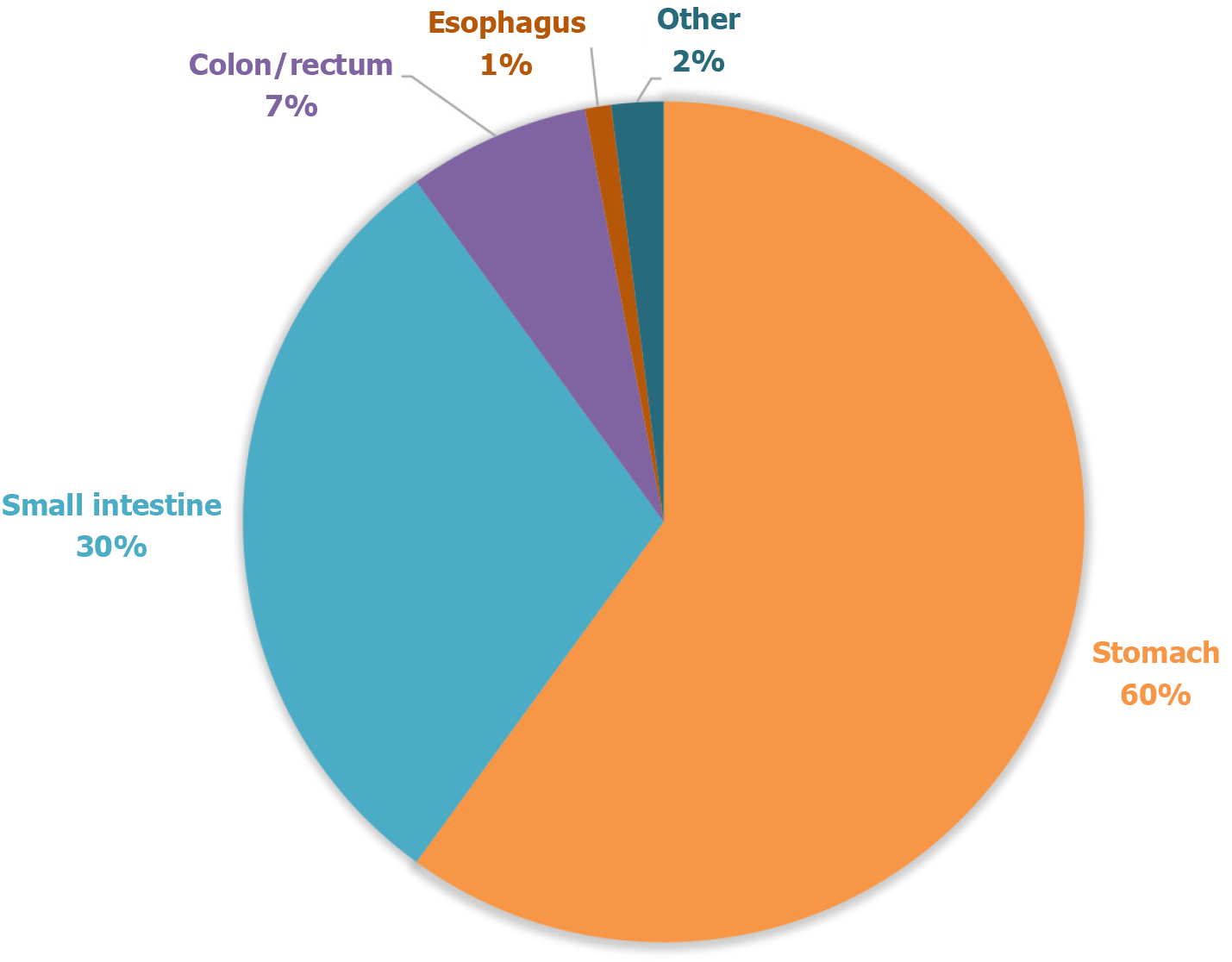
Figure 1 Distribution of gastrointestinal stromal tumors by anatomical location.
The majority of gastrointestinal stromal tumors originate in the stomach (60%) and small intestine (30%), while the esophagus accounts for less than 1% of cases, highlighting the rarity of this localization. Data based on aggregated findings from retrospective clinical studies.
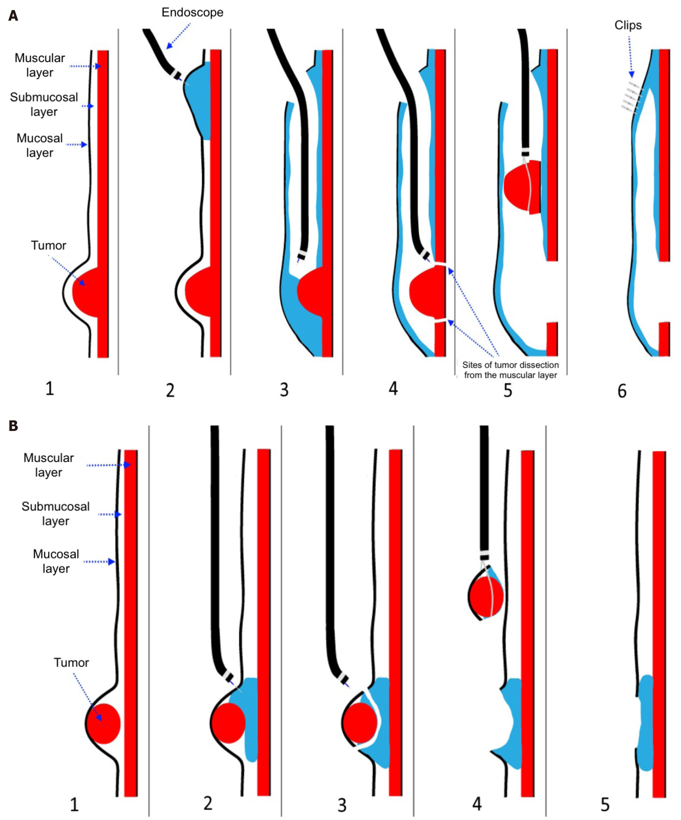
Figure 2 Technique of submucosal tunneling endoscopic resection[18].
A: Technique of submucosal tunneling endoscopic resection: (1) Tumor associated with the muscular membrane of the esophagus; (2) Injection of saline with indigo carmine into the submucosal layer for the safety of initiating slit and forming of submucosal tunnel; (3) Formation of the submucosal tunnel; (4) Enucleation of the tumor; (5) Tumor removal from the submucosal tunnel using a loop; and (6) Closure of the mucosal defect using endoscopic clips; B: Technique of endoscopic full-thickness resection: (1) Tumor associated with the mucous membrane of the esophagus; (2) Injection of saline with indigo carmine into the submucosal layer for safe dissection; (3) Dissection of the tumor; (4) Tumor removal from the esophageal lumen using a loop; and (5) Mucosal defect after tumor dissection. Citation: Smirnov AA, Burakov AN, Blinov EV, Saadulaeva MM, Semenikhin KD, Prudnikov AV, Dvoretskii SI, Kiriltseva MM, Bagnenko SV. The experience of endoscopic resection of benign tumors of the esophagus. Grekov's Bull Surg 2018; 177: 40-44. Copyright ©The Authors 2018. Published by Izdatel'stvo Meditsina Publishers. The authors have obtained the permission (Supplementary material).
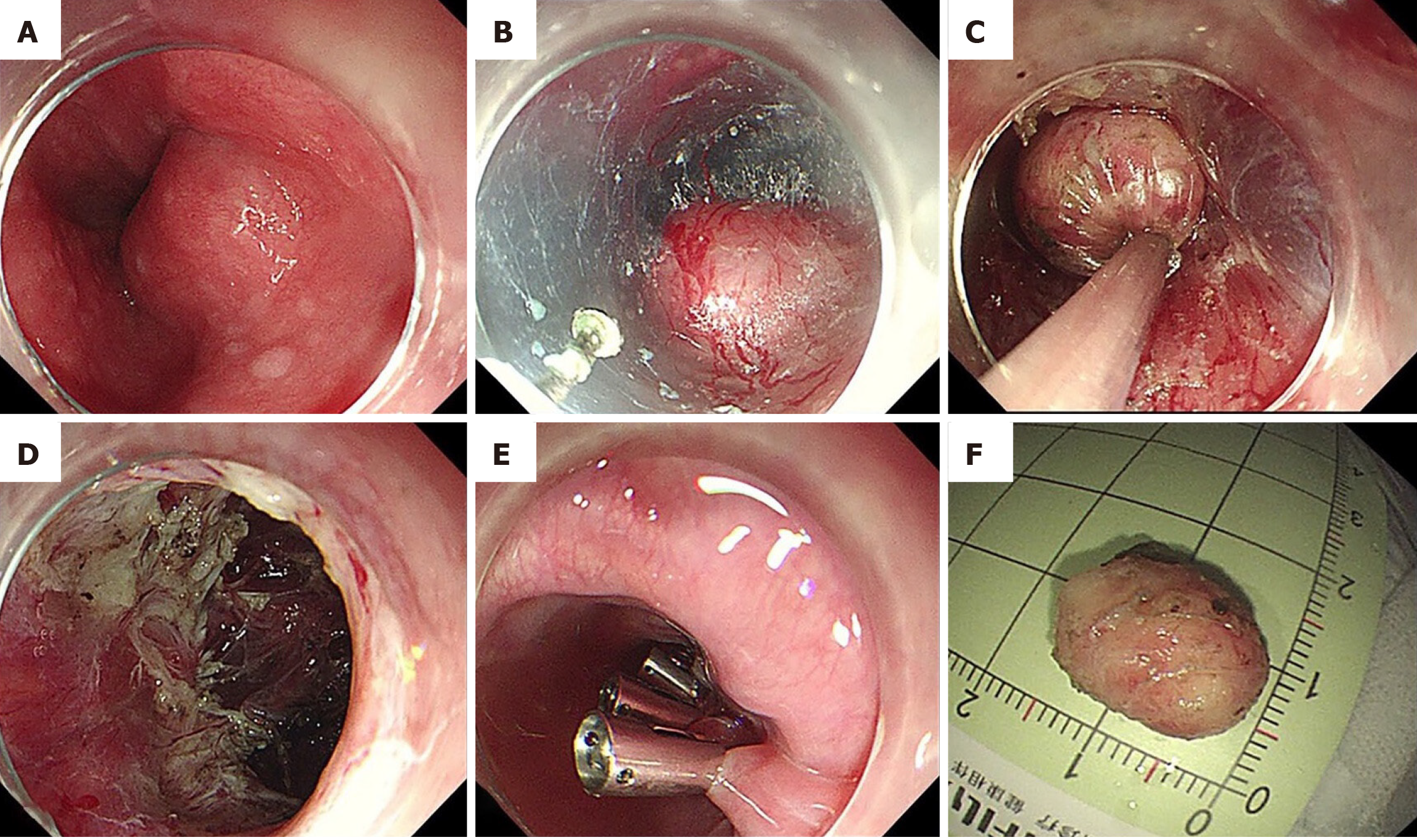
Figure 3 Submucosal tunneling endoscopic resection for a typical esophageal gastrointestinal stromal tumor[19].
A: Endoscopic view of the tumor; B: The tumor was exposed after establishing the mucosal entry established; C: Removal of the resected tumor after complete resection; D: Endoscopic view of the submucosal tunnel after the tumor was removed; E: Incision site was closed by clips; F: Resected specimen. Citation: Lian JJ, Ji YJ, Chen T, Wang GX, Wang MZ, Li SX, Cao J, Shen L, Lu W, Xu MD. Endoscopic resection for esophageal gastrointestinal stromal tumors: A multi-center feasibility study. Ther Adv Gastroenterol 2024; 17: 17562848241255304. Copyright ©The Authors 2024. Published by SAGE Publications. The authors have obtained the permission (Supplementary material).
- Citation: Semash K, Dzhanbekov T. Redefining the treatment paradigm for esophageal gastrointestinal stromal tumors: The emerging role of endoscopic resection. World J Gastroenterol 2025; 31(24): 106440
- URL: https://www.wjgnet.com/1007-9327/full/v31/i24/106440.htm
- DOI: https://dx.doi.org/10.3748/wjg.v31.i24.106440


