Copyright
©The Author(s) 2021.
World J Gastroenterol. Mar 7, 2021; 27(9): 815-834
Published online Mar 7, 2021. doi: 10.3748/wjg.v27.i9.815
Published online Mar 7, 2021. doi: 10.3748/wjg.v27.i9.815
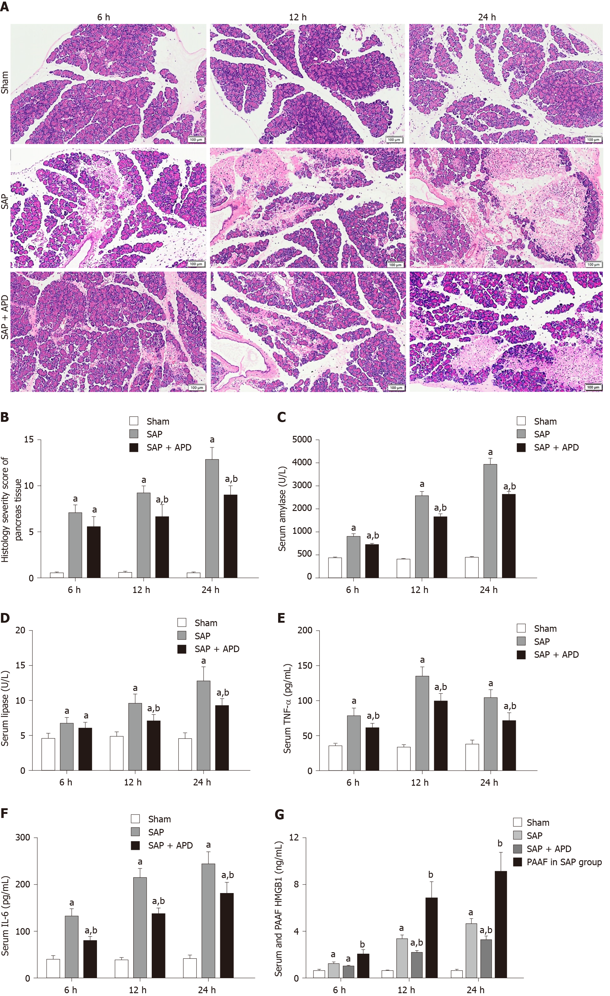
Figure 1 Effects of abdominal paracentesis drainage on pancreatic histopathology and proinflammatory cytokines.
A: Representative images of rat pancreatic tissue from different groups following hematoxylin and eosin staining (bar = 100 μm); B: Histology severity score of pancreas tissue; C: Amylase; D: Lipase; E: Tumor necrosis factor-α; F: Interleukin-6; G: High mobility group box 1. All data are presented as mean ± SD (n = 6). aP < 0.05 vs sham group; bP < 0.05 vs severe acute pancreatitis group. SAP: Severe acute pancreatitis; APD: Abdominal paracentesis drainage; TNF-α: Tumor necrosis factor-α; IL-6: Interleukin-6; PAAF: Pancreatitis-associated ascitic fluid; HMGB1: High mobility group box 1.
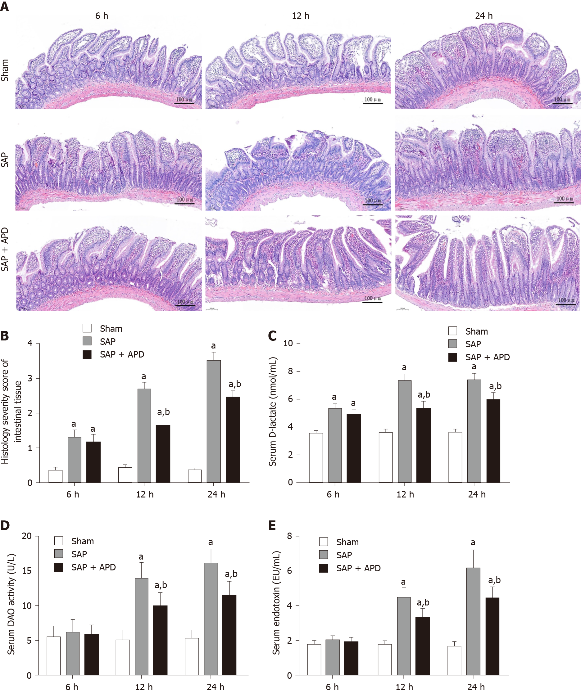
Figure 2 Effects of abdominal paracentesis drainage on intestinal mucosa injury induced by severe acute pancreatitis.
A: Representative images of rat distal ileal tissue from different groups following hematoxylin and eosin staining (bar = 100 μm); B: Histological severity score of intestinal tissues; C: D-lactate; D: Diamine oxidase; E: Endotoxin. All data are presented as mean ± SD (n = 6). aP < 0.05 vs sham group; bP < 0.05 vs severe acute pancreatitis group. SAP: Severe acute pancreatitis; APD: Abdominal paracentesis drainage; DAO; Diamine oxidase.
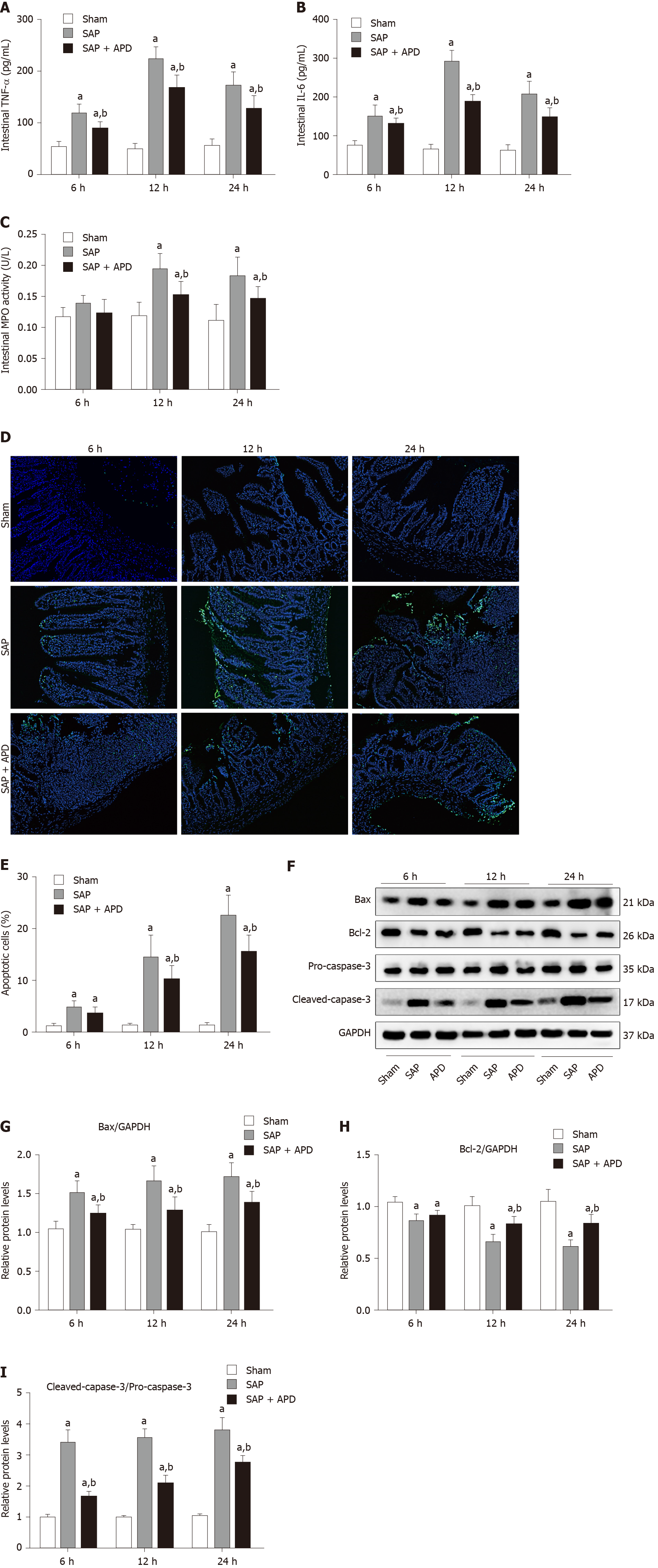
Figure 3 Effects of abdominal paracentesis drainage on intestinal inflammation and mucosal cell apoptosis induced by severe acute pancreatitis.
A: Intestinal tumor necrosis factor-α; B: Intestinal interleukin-6; C: Intestinal myeloperoxidase activity; D: Representative images (400 × magnification) of terminal deoxynucleotidyl-transferase-mediated dUTP nick end labeling (TUNEL) assay; E: Statistical results of the number of apoptotic intestinal mucosal cells in each group; F: Immunoblot of Bax, Bcl-2, pro-caspase-3 and cleaved-caspase-3 protein expression from intestinal samples; G-I: Densitometry analysis of the immunoblot data of apoptosis-related proteins in intestinal tissue. Data are expressed as means ± SD, n = 6 enzyme-linked immunosorbent assay and TUNEL assay results per group; means ± SD, n = 3 immunoblot results per group. aP < 0.05 vs sham group; bP < 0.05 vs severe acute pancreatitis group. TNF-α: Tumor necrosis factor-α; IL-6: Interleukin-6; MPO: Myeloperoxidase; SAP: Severe acute pancreatitis; APD: Abdominal paracentesis drainage; GAPDH: Glyceraldehyde-3-phosphate dehydrogenase.
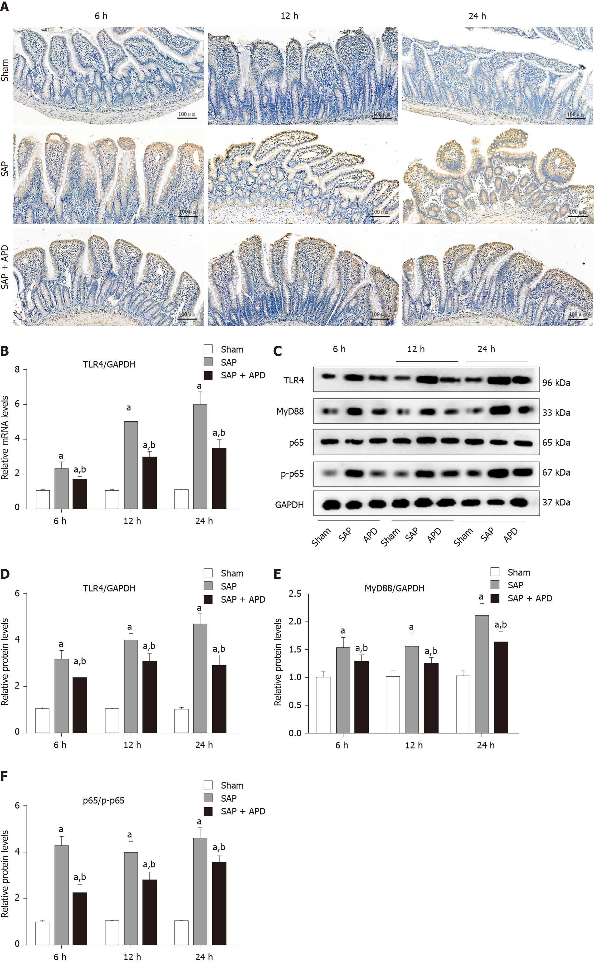
Figure 4 Effects of abdominal paracentesis drainage on intestinal toll-like receptor 4 signaling pathway in rats with severe acute pancreatitis.
A: Representative immunohistochemistry images of toll-like receptor 4 (TLR4) in intestinal tissue (bar = 100 μm); B: TLR4 mRNA measurement by real-time polymerase chain reaction (PCR); C: Immunoblotting of TLR4, myeloid differentiation factor 88 (MyD88) and p65 protein expression and p65 phosphorylation level from intestinal samples; D-F: Densitometry analysis of TLR4, MyD88 and ratio of p65 to phosphorylated p65. Data are expressed as means ± SD, n = 6 real-time PCR results per group; means ± SD, n = 3 immunoblotting results per group. aP < 0.05 vs sham group; bP < 0.05 vs severe acute pancreatitis group. SAP: Severe acute pancreatitis; APD: Abdominal paracentesis drainage; MyD88: Myeloid differentiation factor 88; TLR4: Toll-like receptor 4; p-p65: Phosphorylated p65; GAPDH: Glyceraldehyde-3-phosphate dehydrogenase.
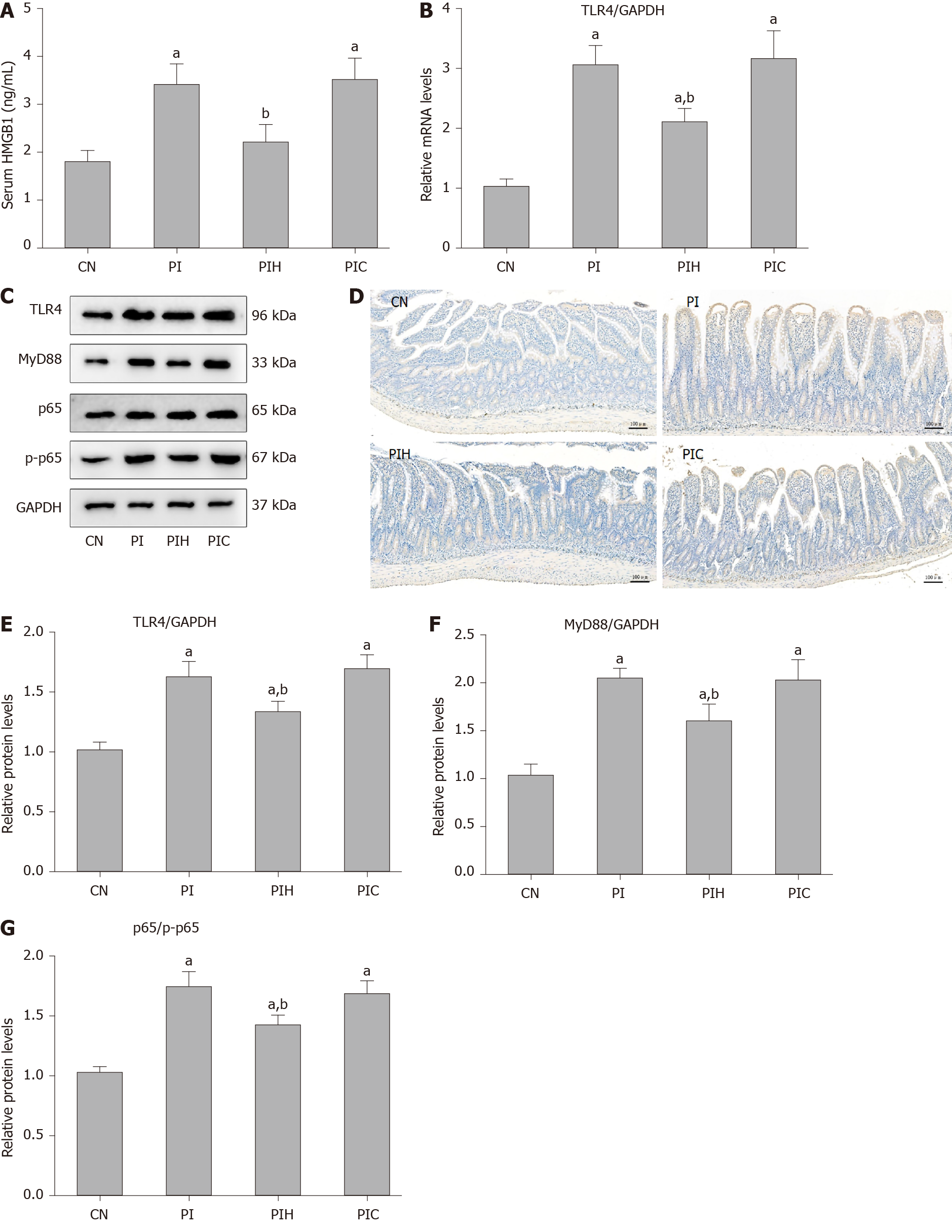
Figure 5 Effects of pancreatitis-associated ascitic fluid intraperitoneal injection with or without anti-high mobility group box protein 1 neutralizing antibody on intestinal toll-like receptor 4 signaling pathway in cerulein-treated rats.
A: High mobility group box protein 1 in serum; B: Toll-like receptor 4 (TLR4) mRNA measurement by real-time polymerase chain reaction; C: Immunoblotting of TLR4, myeloid differentiation factor 88 (MyD88) and p65 protein expression and p65 phosphorylation level from intestine samples; D: Representative immunohistochemical images of TLR4 in intestinal tissue (bar = 100 μm); E-G: Densitometry analysis of TLR4, MyD88 and ratio of p65 to phosphorylated p65. Data are expressed as means ± SD, n = 6 enzyme-linked immunosorbent assay results per group; means ± SD, n = 3 immunoblotting results per group. aP < 0.05 vs control group; bP < 0.05 vs pancreatitis-associated ascitic fluid injection group. HMGB1: High mobility group box protein 1; CN: Control; PI: Pancreatitis-associated ascitic fluid (PAAF) injection; PIH: PAAF and 200 μg anti-HMGB1 neutralizing antibody; PIC: PAAF + control lgY; TLR4: Toll-like receptor 4; MyD88: Myeloid differentiation factor 88; p-p65: Phosphorylated p65; GAPDH: Glyceraldehyde-3-phosphate dehydrogenase.
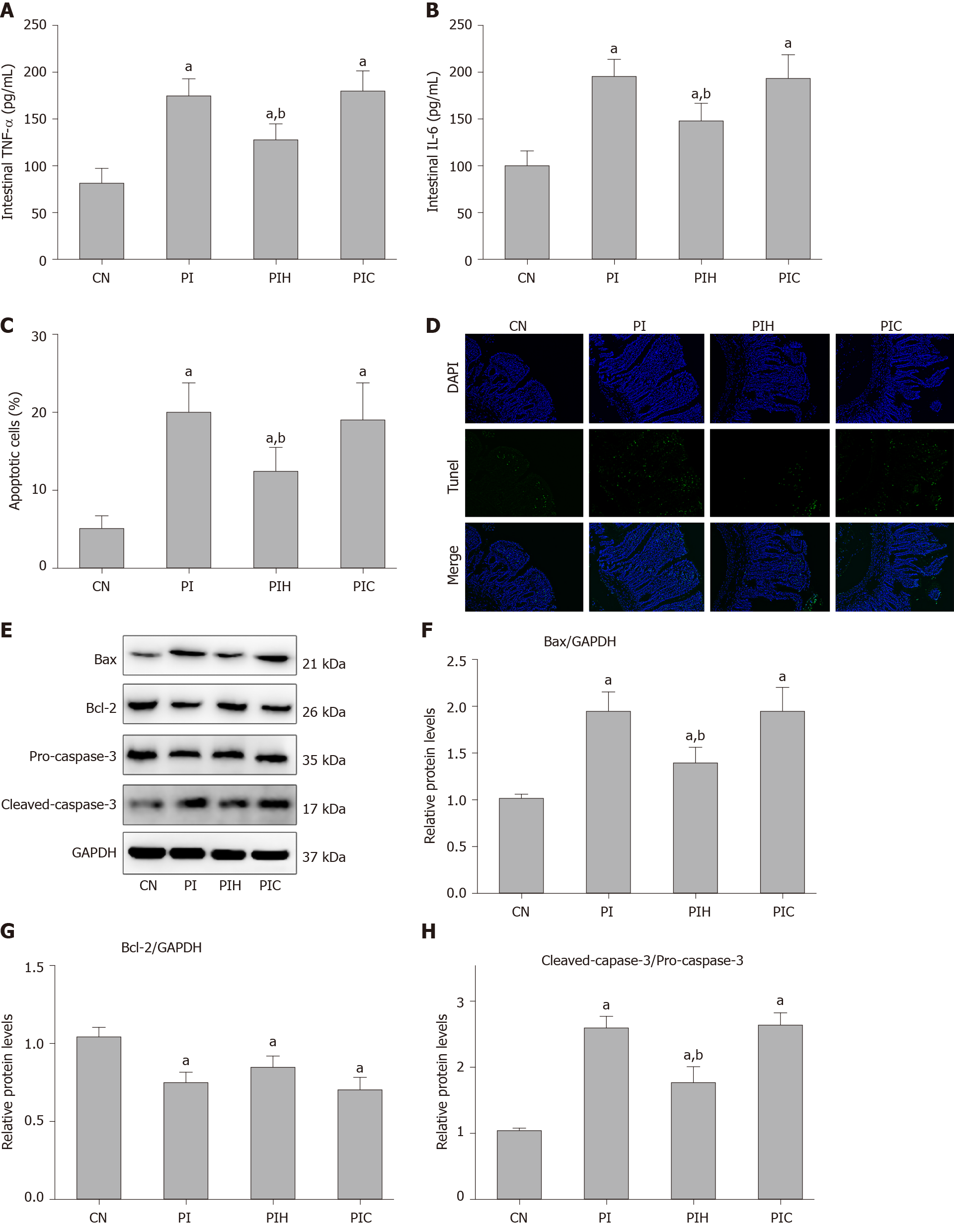
Figure 6 Effects of intraperitoneal pancreatitis-associated ascitic fluid injection with or without anti-high mobility group box protein 1 neutralizing antibody on intestinal inflammation and cell apoptosis in cerulein-treated rats.
A: Intestinal tumor necrosis factor-α; B: Intestinal interleukin-6; C: Statistical results of the number of apoptotic intestinal mucosal cells in each group; D: Representative images (400 × magnification) of terminal deoxynucleotidyl-transferase-mediated dUTP nick end labeling (TUNEL) assay; E: Immunoblotting of Bax, Bcl-2, pro-caspase-3 and cleaved-caspase-3 protein expression from intestinal samples; F-H: Densitometry analysis of the immunoblotting data of apoptosis-related proteins in intestinal tissue. Data are expressed as means ± SD, n = 6 enzyme-linked immunosorbent assay and TUNEL assay results per group; means ± SD, n = 3 immunoblotting results per group. aP < 0.05 vs control group; bP < 0.05 vs pancreatitis-associated ascitic fluid group. TNF-α: Tumor necrosis factor-α; CN: Control; PI: Pancreatitis-associated ascitic fluid (PAAF) injection; PIH: PAAF and 200 μg anti-high mobility group box protein 1 neutralizing antibody; PIC: PAAF + control lgY; IL-6: Interleukin-6; TUNEL: Terminal deoxynucleotidyl-transferase-mediated dUTP nick end labeling; GAPDH: Glyceraldehyde-3-phosphate dehydrogenase; DAPI: 4',6-diamidino-2-phenylindole.
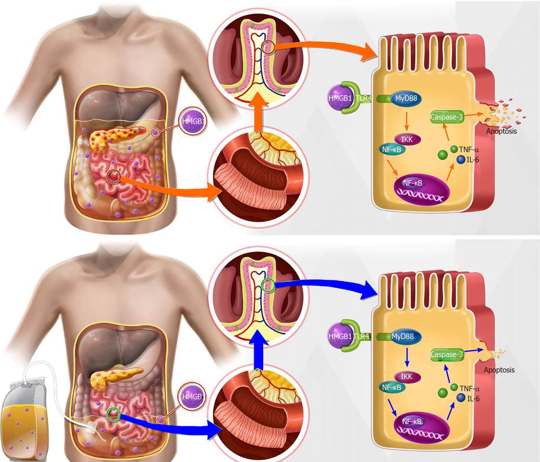
Figure 7 Possible mechanisms responsible for the protective effects of abdominal paracentesis drainage treatment on severe acute pancreatitis-induced intestinal inflammation and accompanying apoptosis.
TNF-α: Tumor necrosis factor-α; IL-6: Interleukin-6; HMGB1: High mobility group box protein 1; IKK: IkappaB kinase; NF-kB: Nuclear factor kB; TLR4: Toll-like receptor 4.
- Citation: Huang SQ, Wen Y, Sun HY, Deng J, Zhang YL, Huang QL, Wang B, Luo ZL, Tang LJ. Abdominal paracentesis drainage attenuates intestinal inflammation in rats with severe acute pancreatitis by inhibiting the HMGB1-mediated TLR4 signaling pathway. World J Gastroenterol 2021; 27(9): 815-834
- URL: https://www.wjgnet.com/1007-9327/full/v27/i9/815.htm
- DOI: https://dx.doi.org/10.3748/wjg.v27.i9.815


