Copyright
©The Author(s) 2020.
World J Gastroenterol. Dec 14, 2020; 26(46): 7425-7435
Published online Dec 14, 2020. doi: 10.3748/wjg.v26.i46.7425
Published online Dec 14, 2020. doi: 10.3748/wjg.v26.i46.7425
Figure 1 Periorbital and facial edema upon the patient’s diagnosis of dermatomyositis in 2016.
Figure 2 Pathology of left upper arm muscle.
Transverse striations of muscle fibers can be seen. Atrophy and fibrosis were not observed. Local intermuscular vascular dilatation and infiltration with a small amount of lymphocytes were noted.
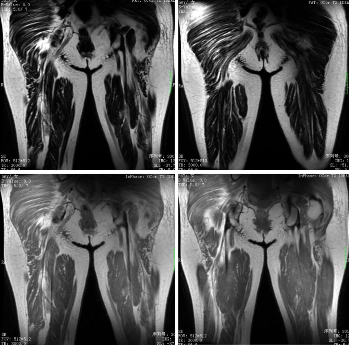
Figure 3 Plain magnetic resonance imaging scan of the middle and upper parts of the right thigh.
A patchy higher signal can be seen in the fat-inhibited part of the T2-weighted images of multiple muscle tissues in buttocks and middle and upper thigh with unclear boundary and uneven signal.
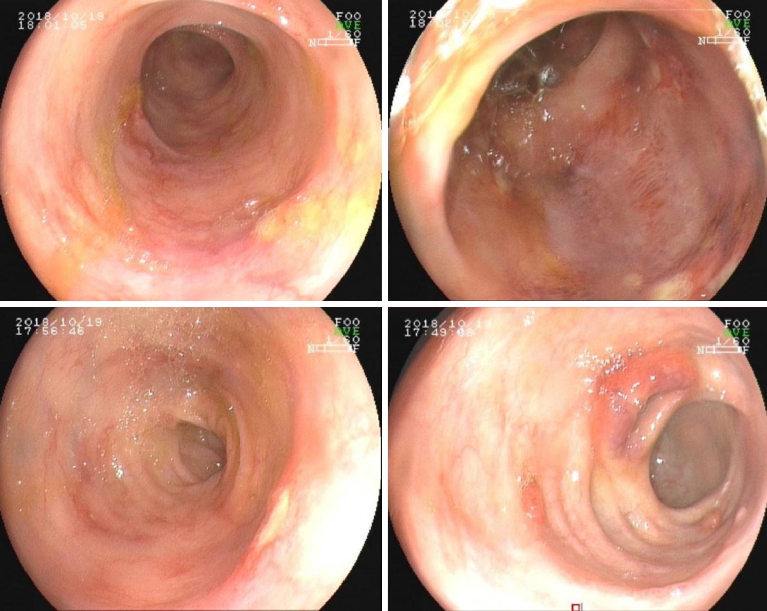
Figure 4 Colonoscopic presentation of ulcerative colitis.
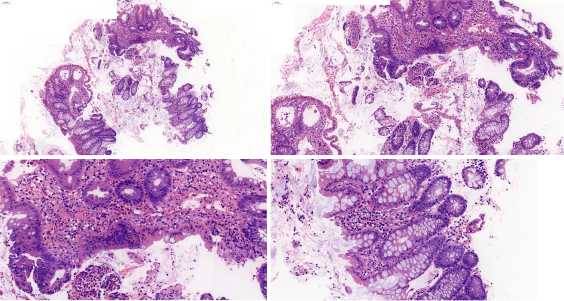
Figure 5 Intestinal mucosal biopsy at the time of diagnosis of ulcerative colitis.
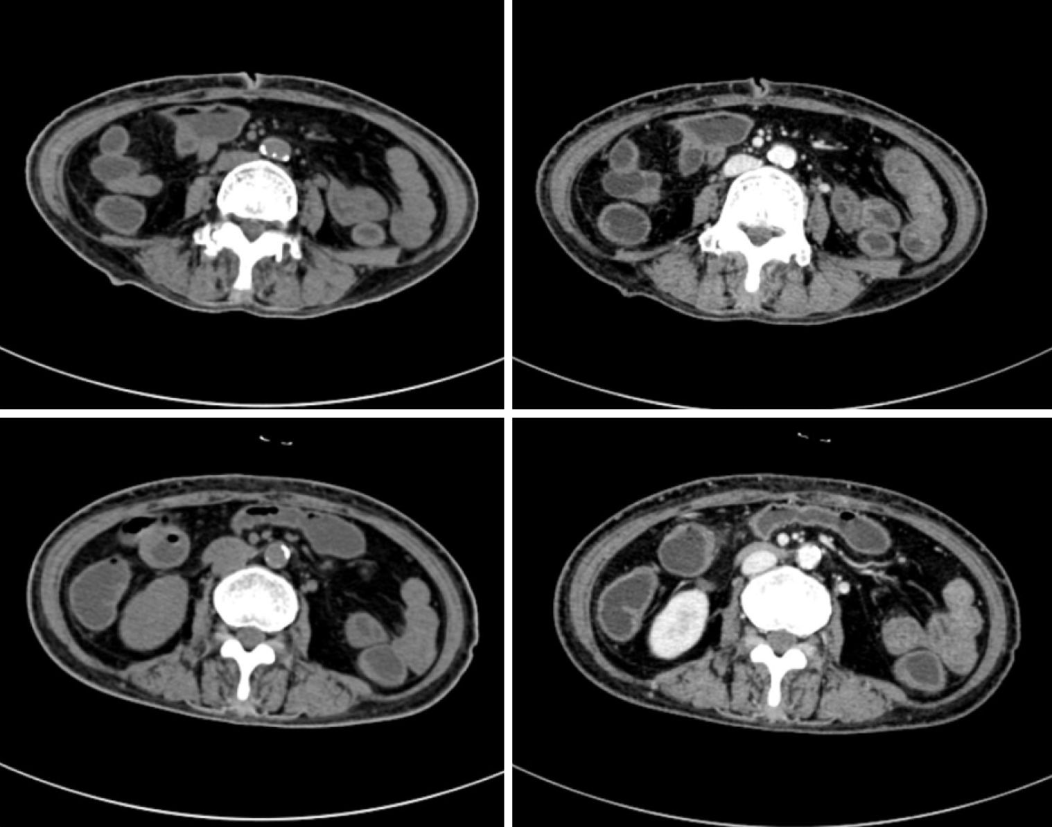
Figure 6 Abdominal computed tomography at the time of diagnosis of ulcerative colitis.
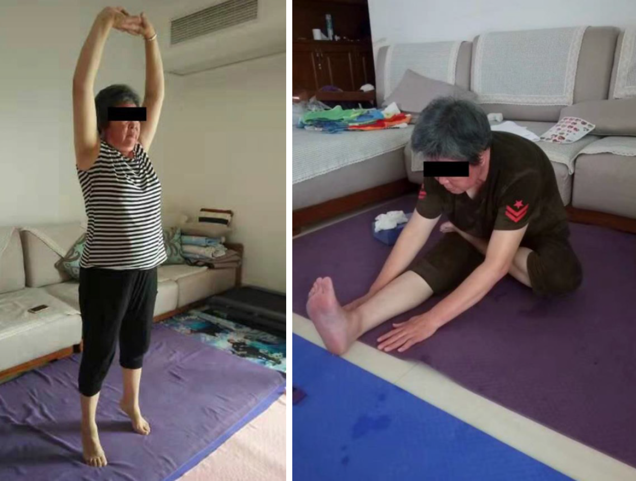
Figure 7 Patient status after treatment with infliximab.
The patient recovered her muscle strength in proximal extremities and could freely move without needing a wheelchair.
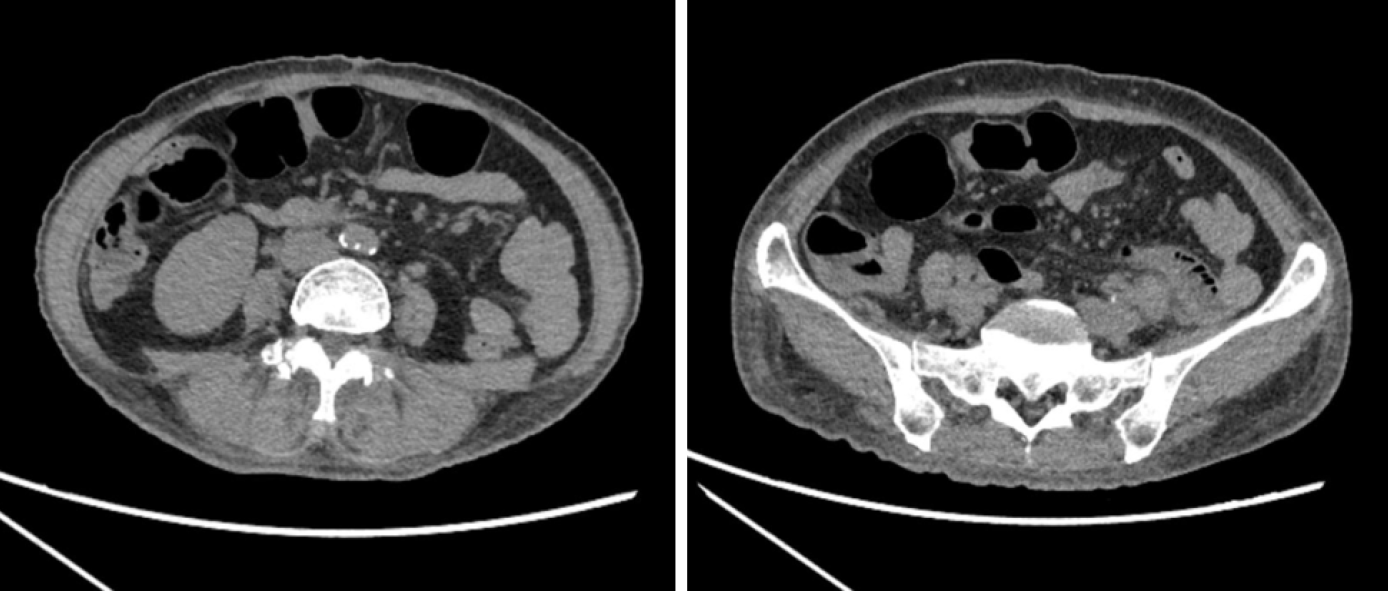
Figure 8 Computed tomography re-examination of the small intestines after the fourth infliximab treatment.
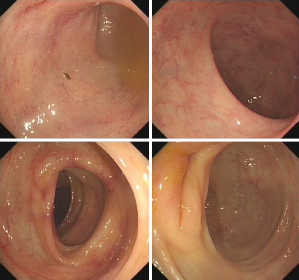
Figure 9 Colonoscopy re-examination after the fourth infliximab treatment.
- Citation: Huang BB, Han LC, Liu GF, Lv XD, Gu GL, Li SQ, Chen L, Wang HQ, Zhan LL, Lv XP. Infliximab is effective in the treatment of ulcerative colitis with dermatomyositis: A case report. World J Gastroenterol 2020; 26(46): 7425-7435
- URL: https://www.wjgnet.com/1007-9327/full/v26/i46/7425.htm
- DOI: https://dx.doi.org/10.3748/wjg.v26.i46.7425


