Copyright
©The Author(s) 2020.
World J Gastroenterol. Dec 14, 2020; 26(46): 7312-7324
Published online Dec 14, 2020. doi: 10.3748/wjg.v26.i46.7312
Published online Dec 14, 2020. doi: 10.3748/wjg.v26.i46.7312
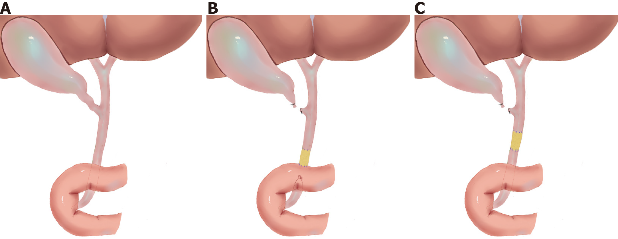
Figure 1 Diagrams of common bile ductal injury and repair model.
A: Normal extrahepatic common bile duct; B: Injury and repair model of distal common bile duct (group A); C: Injury and repair model of intermedial common bile duct (group B).
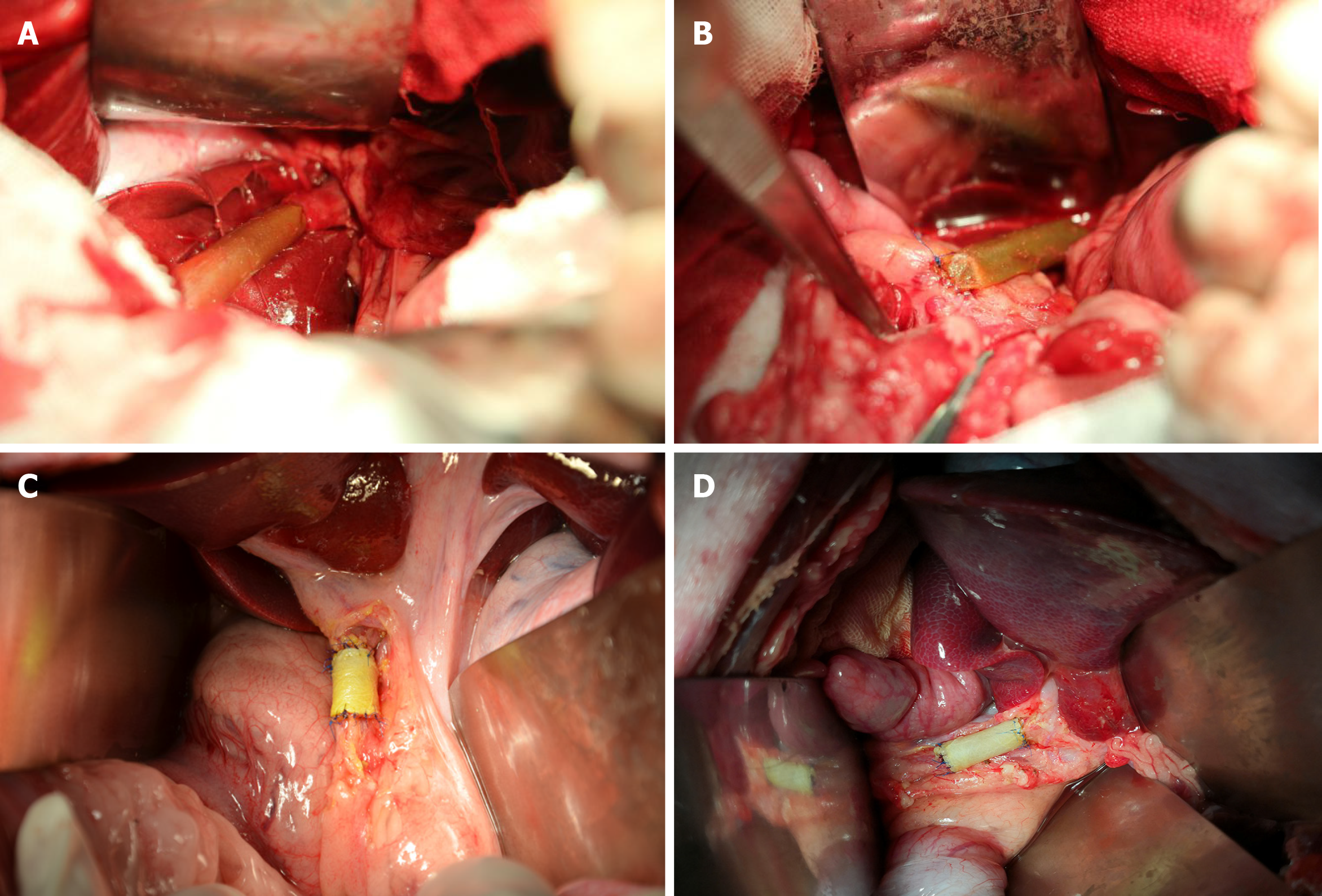
Figure 2 Surgical procedures of extrahepatic common bile duct reconstruction.
A: End-to-end anastomosis was carried out between the proximal stump of the common bile duct (CBD) and the animal-derived artificial bile duct (2 cm in length) after resection of a 2 cm distal CBD segment (group A); B: End-to-side anastomosis was performed between the distal stump of the animal-derived artificial bile duct and the duodenum to complete the repairment of the distal CBD defect (group A); C and D: End-to-end anastomoses were conducted between the stumps of CBD and the animal-derived artificial bile duct (2 cm in length) to repair the 2 cm intermedial CBD defect (group B).
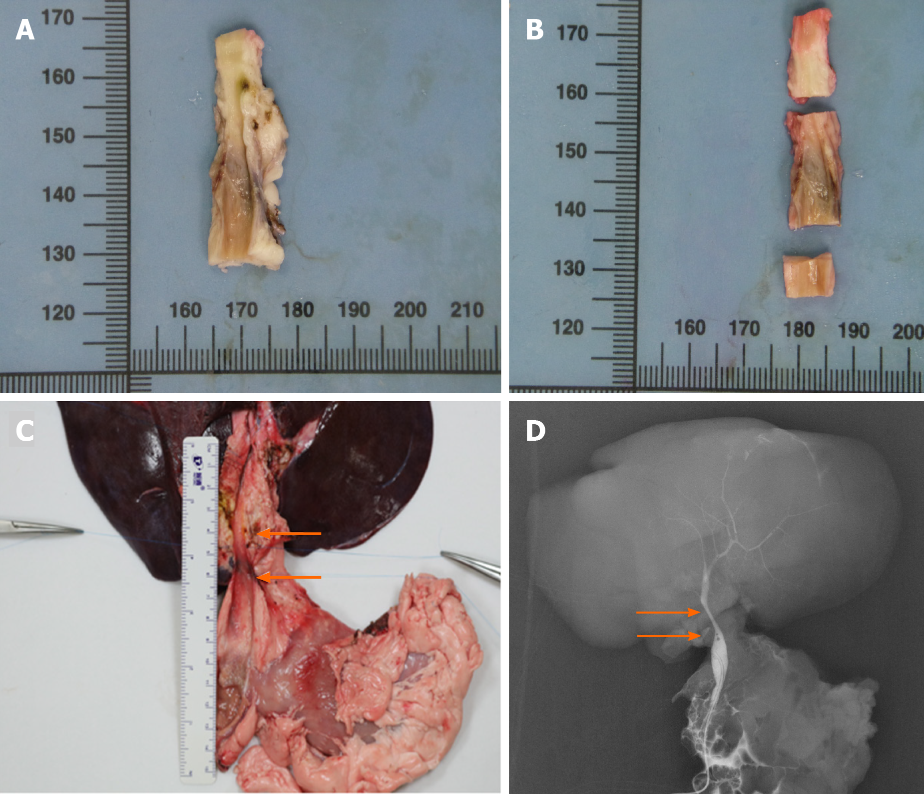
Figure 3 Autopsy and cholangiography.
A: The macroscopic assessment of the extrahepatic bile duct (BD) indicated the inner walls of the anastomoses were smooth without scars or stricture; B: The extrahepatic BD was divided into three segments to perform the microscopic assessment according to the anastomoses; C: The en bloc liver procured was associated with the entire extrahepatic BD including partial duodenum and pancreas after euthanasia. The area of two white arrows denoted the location of the animal-derived artificial bile duct (group B); D: The cholangiography showed patency of the animal-derived artificial bile duct without dilatation of the proximal BD. The area of two white arrows denoted the location of the animal-derived artificial bile duct (group B).
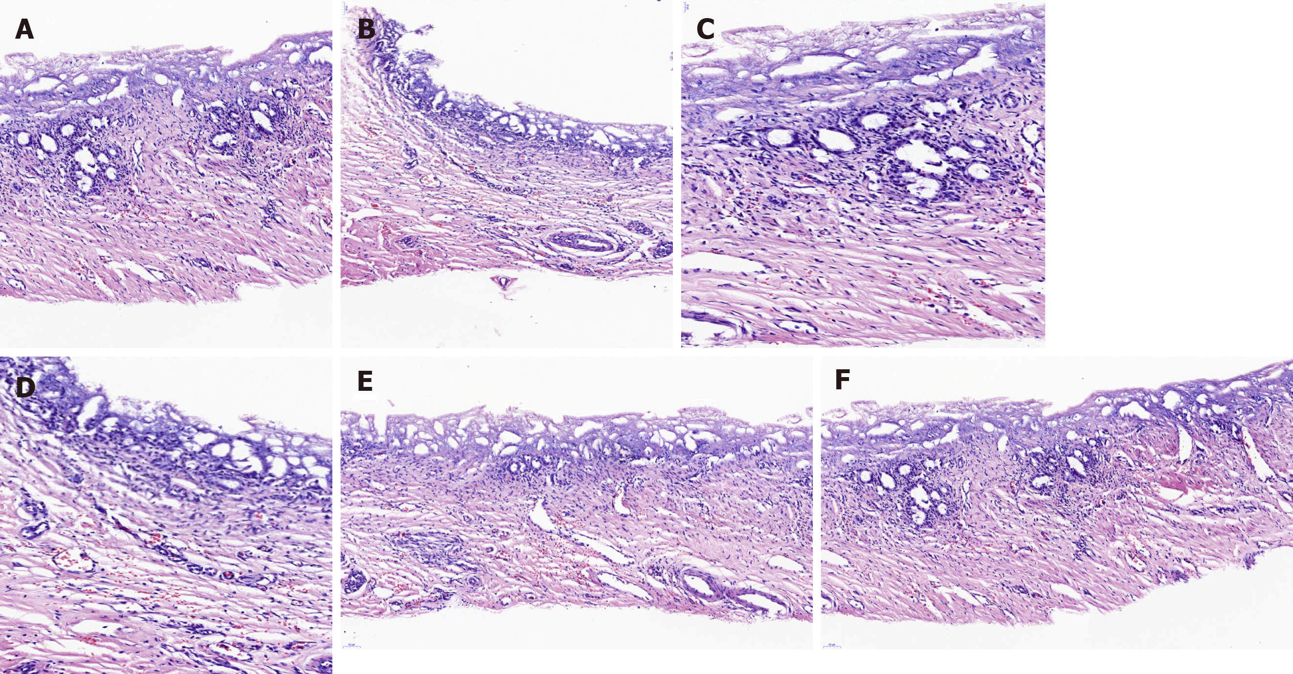
Figure 4 Hematoxylin/eosin staining of the animal-derived artificial bile duct site.
No marked transmural neutrophil and lymphoplasma cell infiltration was observed. A: Hematoxylin/eosin (H/E) staining of a 6 mo survival pig in group A (magnification: 20 ×); B: H/E staining of a 12 mo survival pig in group B (magnification: 20 ×); C: H/E staining of a 6 mo survival pig in group A (magnification: 40 ×); D: H/E staining of a 12 mo survival pig in group B (magnification: 40 ×); E and F: H/E staining result of neovascularization was marked in group A and B (magnification: 20 ×).
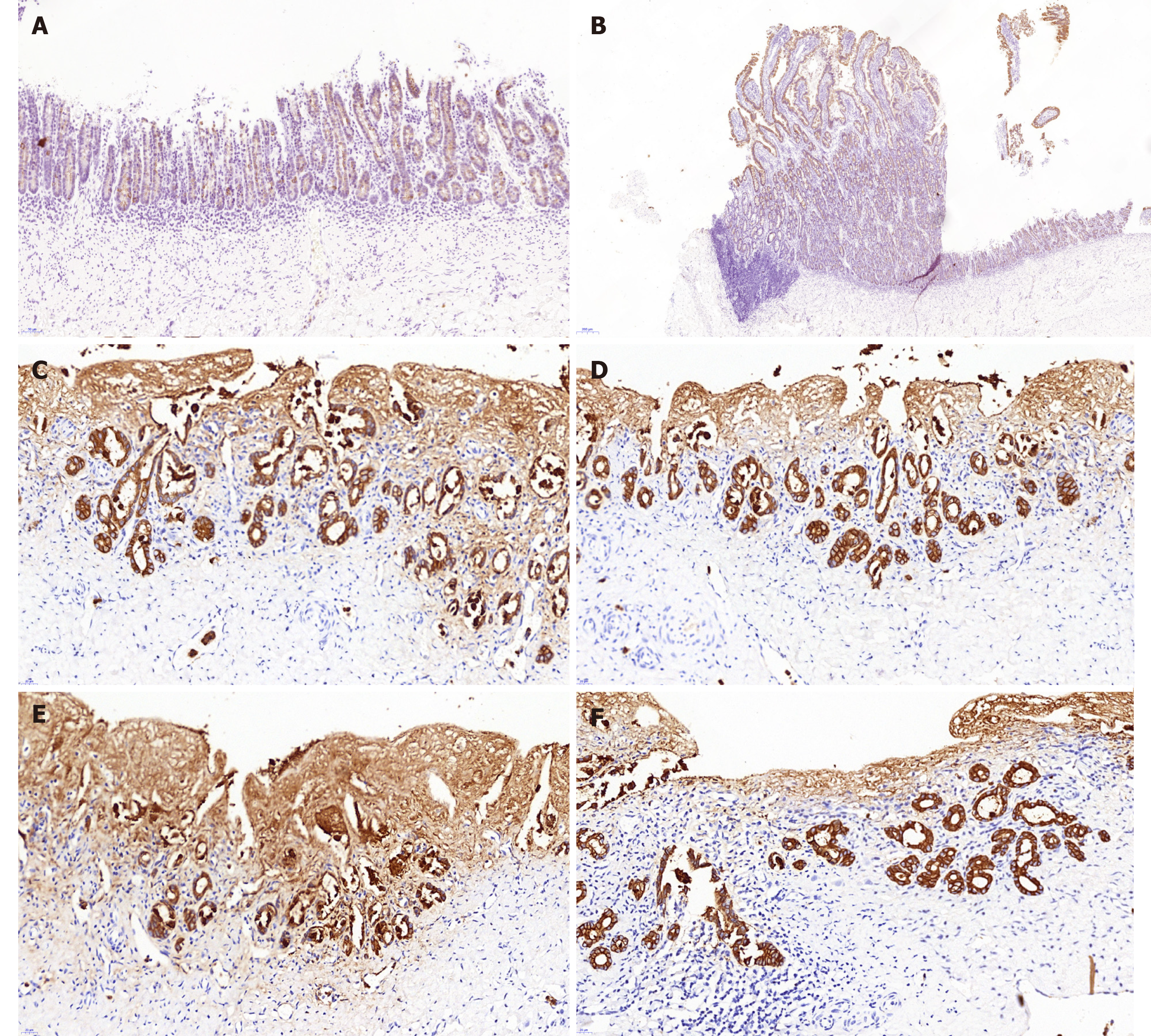
Figure 5 Cytokeratin 19 immunostaining.
The cytokeratin 19 (CK19) staining showed no marked differences between the animal-derived artificial bile duct site and the normal bile duct on the aspect of bile duct epithelium and accessory glands on the inner wall. The superior integrity of regeneration of the biliary epithelial layer was observed in group B than that in group A. A: CK19 staining of the animal-derived artificial bile duct site in group A (magnification: 20 ×); B: CK19 staining of duodenal papilla in group A (magnification: 20 ×); C and D: CK19 staining of the animal-derived artificial bile duct site in group B (magnification: 40 ×); E and F: CK19 staining of the distal normal bile duct in group B (magnification: 40 ×).
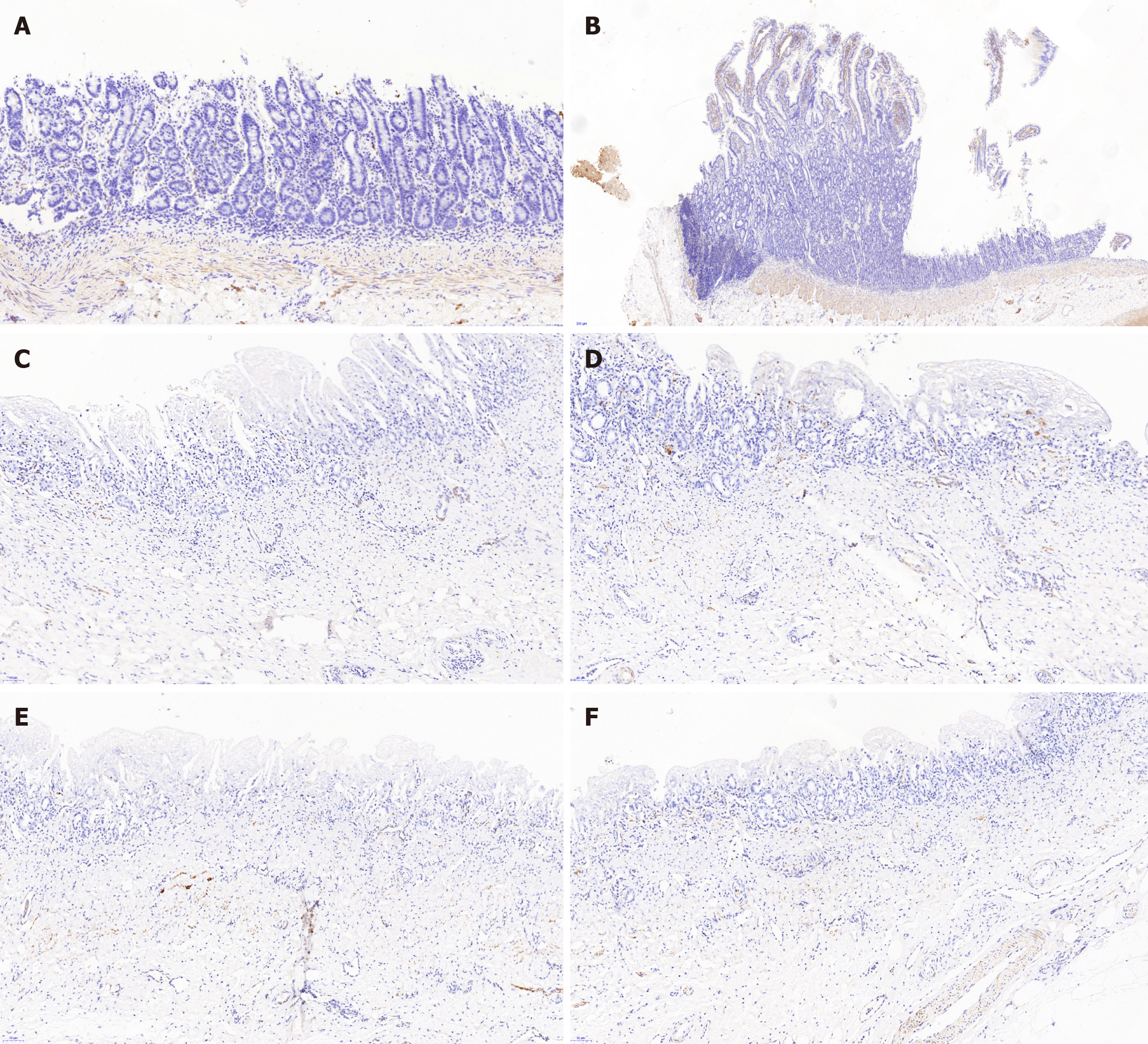
Figure 6 Desmin immunostaining.
The desmin positive area confirmed the regeneration of smooth muscle. Meanwhile, the desmin positive area was significantly larger in group A than group B. A: Desmin immunostaining of the animal-derived artificial bile duct site in group A (magnification: 20 ×); B: Desmin immunostaining of duodenal papilla in group A (magnification: 20 ×); C and D: Desmin immunostaining of the animal-derived artificial bile duct site in group B (magnification: 20 ×); E and F: Desmin immunostaining of the distal normal bile duct in group B (magnification: 20 ×).
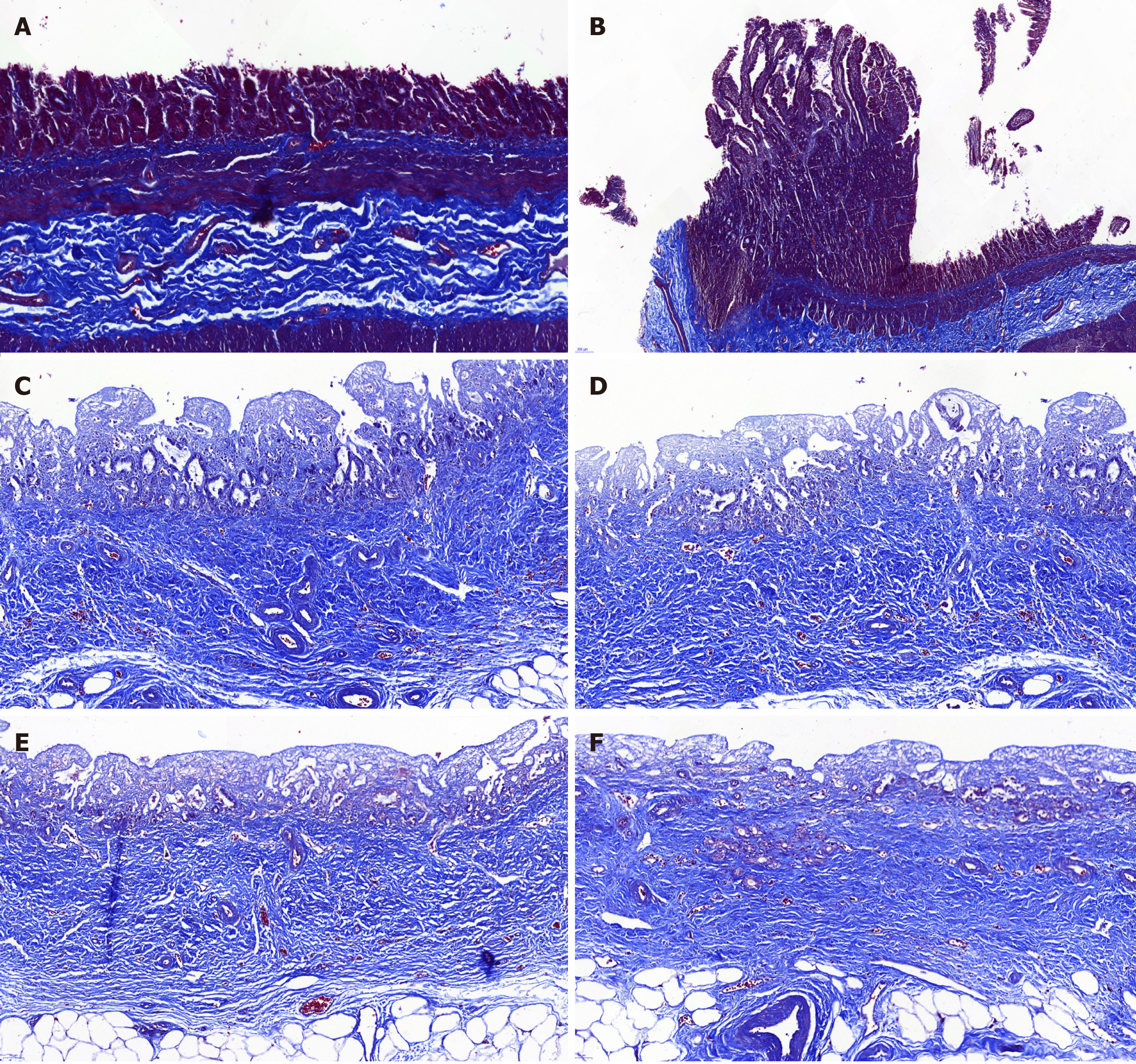
Figure 7 Masson’s trichrome staining.
The Masson’s trichrome staining showed marked regeneration of the submucosal connective tissue and no significant differences between the animal-derived artificial bile duct site and the normal bile duct. The submucosal connective tissue was more regular and thicker in group B than that in group A. A: Masson staining of the animal-derived artificial bile duct site in group A (magnification: 20 ×); B: Masson staining of duodenal papilla in group A (magnification: 20 ×); C and D: Masson staining of the animal-derived artificial bile duct site in group B (magnification: 20 ×); E and F: Masson staining of the distal normal bile duct in group B (magnification: 20 ×).
- Citation: Shang H, Zeng JP, Wang SY, Xiao Y, Yang JH, Yu SQ, Liu XC, Jiang N, Shi XL, Jin S. Extrahepatic bile duct reconstruction in pigs with heterogenous animal-derived artificial bile ducts: A preliminary experience. World J Gastroenterol 2020; 26(46): 7312-7324
- URL: https://www.wjgnet.com/1007-9327/full/v26/i46/7312.htm
- DOI: https://dx.doi.org/10.3748/wjg.v26.i46.7312


