Copyright
©The Author(s) 2020.
World J Gastroenterol. Dec 14, 2020; 26(46): 7312-7324
Published online Dec 14, 2020. doi: 10.3748/wjg.v26.i46.7312
Published online Dec 14, 2020. doi: 10.3748/wjg.v26.i46.7312
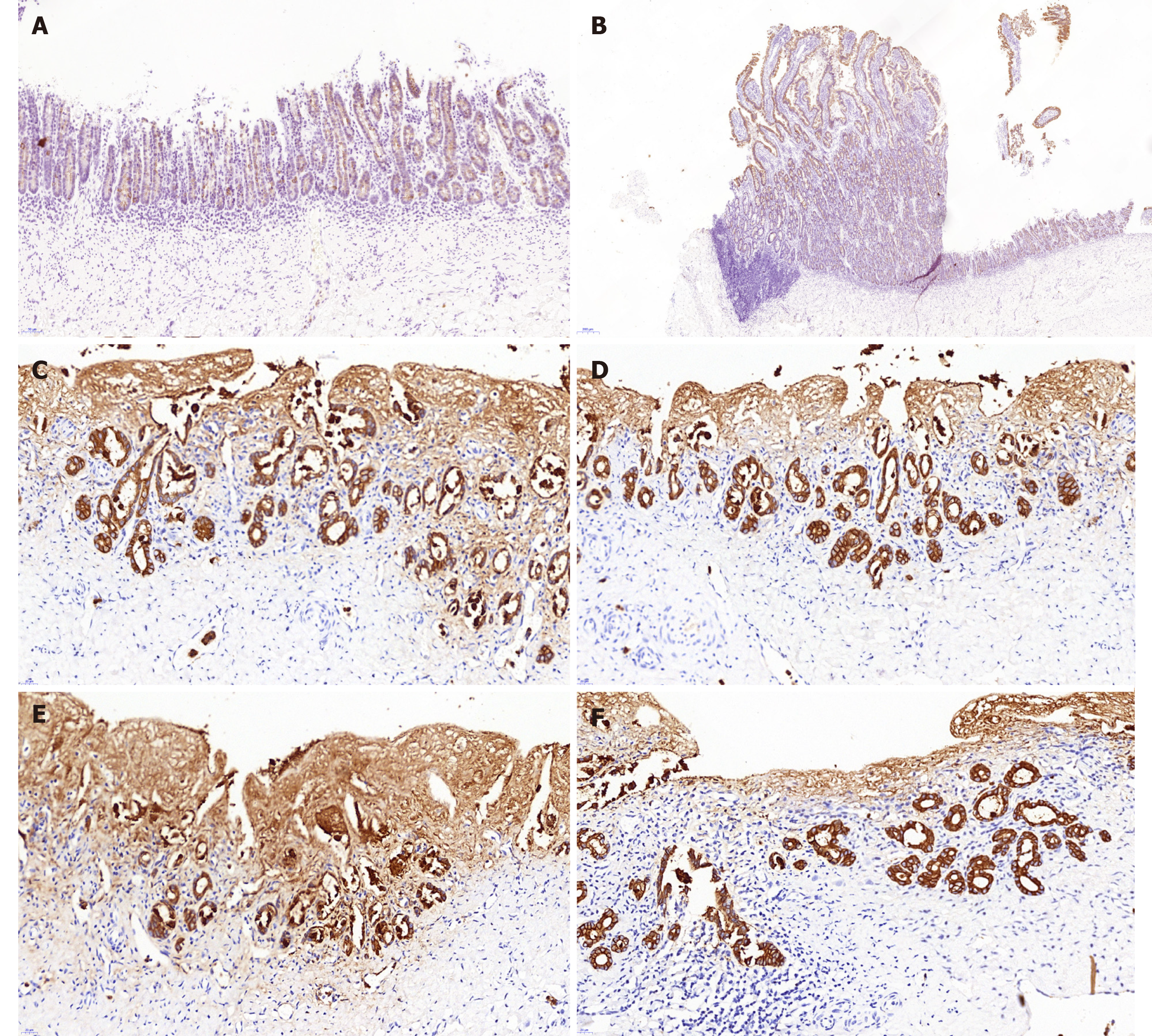
Figure 5 Cytokeratin 19 immunostaining.
The cytokeratin 19 (CK19) staining showed no marked differences between the animal-derived artificial bile duct site and the normal bile duct on the aspect of bile duct epithelium and accessory glands on the inner wall. The superior integrity of regeneration of the biliary epithelial layer was observed in group B than that in group A. A: CK19 staining of the animal-derived artificial bile duct site in group A (magnification: 20 ×); B: CK19 staining of duodenal papilla in group A (magnification: 20 ×); C and D: CK19 staining of the animal-derived artificial bile duct site in group B (magnification: 40 ×); E and F: CK19 staining of the distal normal bile duct in group B (magnification: 40 ×).
- Citation: Shang H, Zeng JP, Wang SY, Xiao Y, Yang JH, Yu SQ, Liu XC, Jiang N, Shi XL, Jin S. Extrahepatic bile duct reconstruction in pigs with heterogenous animal-derived artificial bile ducts: A preliminary experience. World J Gastroenterol 2020; 26(46): 7312-7324
- URL: https://www.wjgnet.com/1007-9327/full/v26/i46/7312.htm
- DOI: https://dx.doi.org/10.3748/wjg.v26.i46.7312


