Copyright
©The Author(s) 2018.
World J Gastroenterol. Jul 14, 2018; 24(26): 2806-2817
Published online Jul 14, 2018. doi: 10.3748/wjg.v24.i26.2806
Published online Jul 14, 2018. doi: 10.3748/wjg.v24.i26.2806
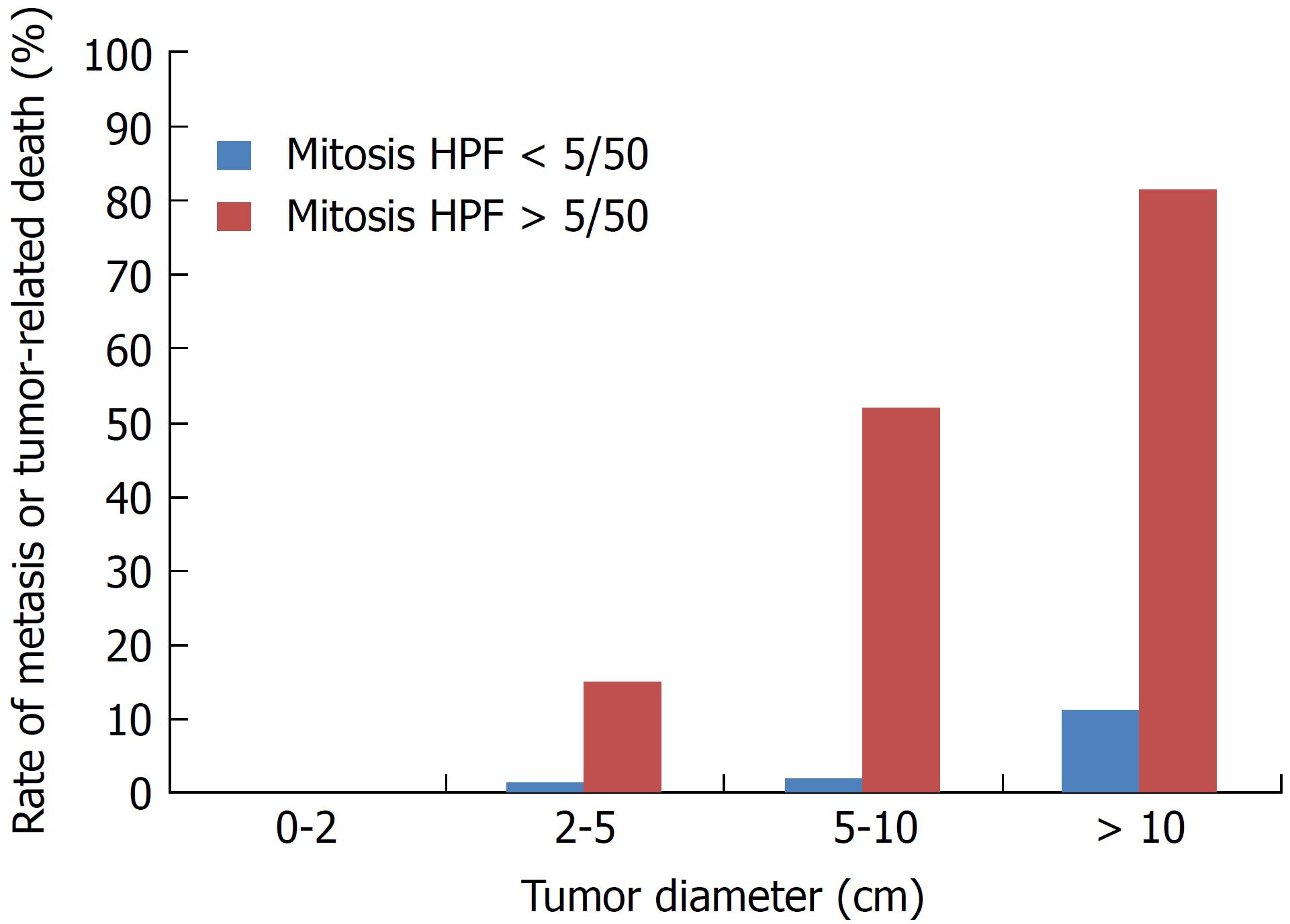
Figure 1 Rate of metastasis or tumor-related death according to tumor diameter and mitotic index.
Created using reference[6].
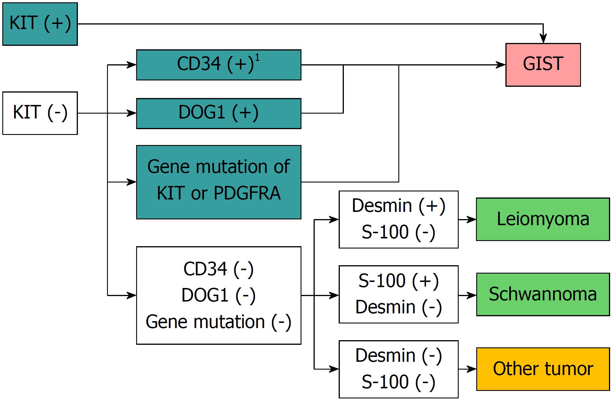
Figure 2 Flow chart of diagnosis of gastrointestinal mesenchymal tumors using immunohistochemical or genetic analysis.
1Solitary fibrous tumors should be ruled out. Quoted and modified from reference[8]. GIST: Gastrointestinal stromal tumor.
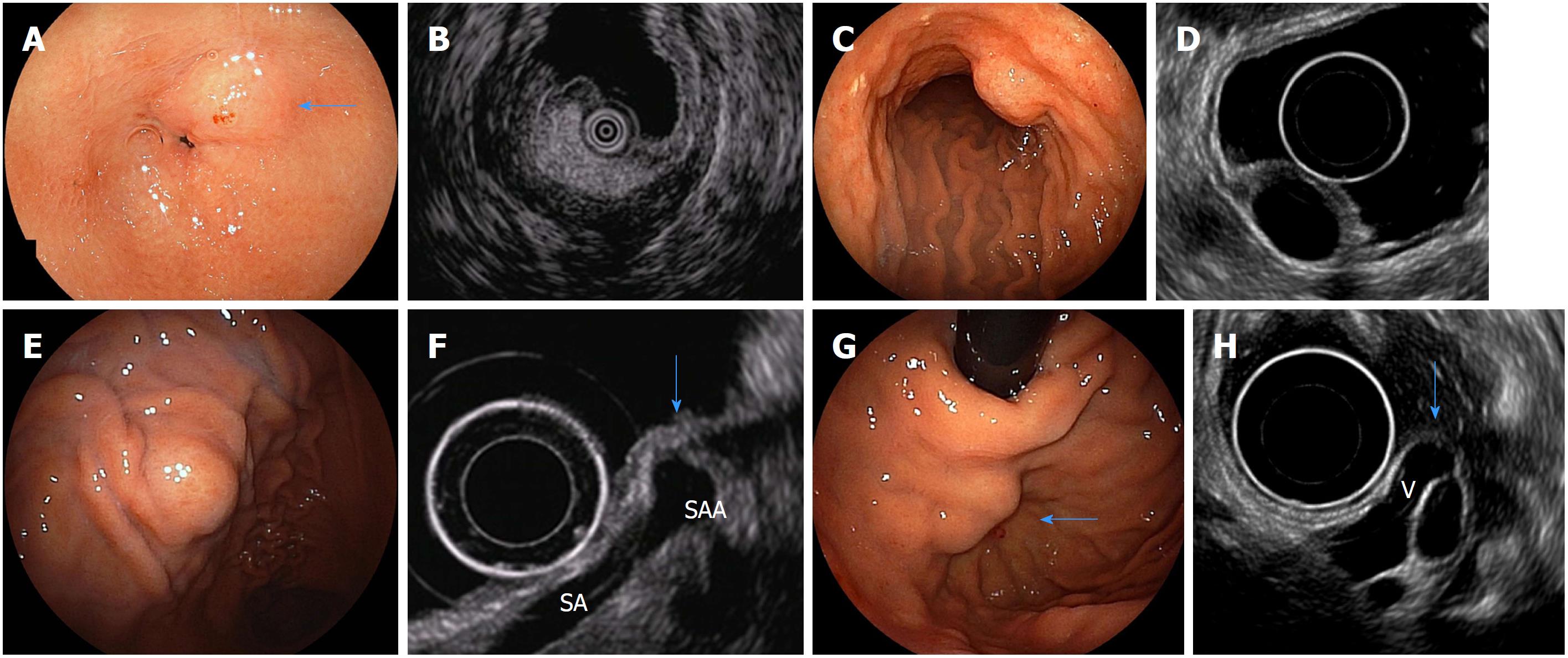
Figure 3 Endoscopic images of subepithelial lesions that can be diagnosed only with endoscopic ultrasound findings and their specific endoscopic ultrasonography images.
A: Endoscopic image of a gastric lipoma (arrow); B: Endoscopic ultrasound (EUS) image of A (high-echo mass); C: Endoscopic image of a gastric cyst; D: EUS image of C (anechoic mass); E: Endoscopic image of extra-gastric compression due to splenic artery aneurysm; F: EUS image of E [normal gastric wall is compressed by a splenic artery aneurysm(SAA) (arrow). SA: splenic artery]; G: Endoscopic image of gastric varices (arrow); H: EUS image of G [varices are present in the submucosa from the outside of the wall (V) (arrow)]. Quoted and modified from reference[38] with permission.
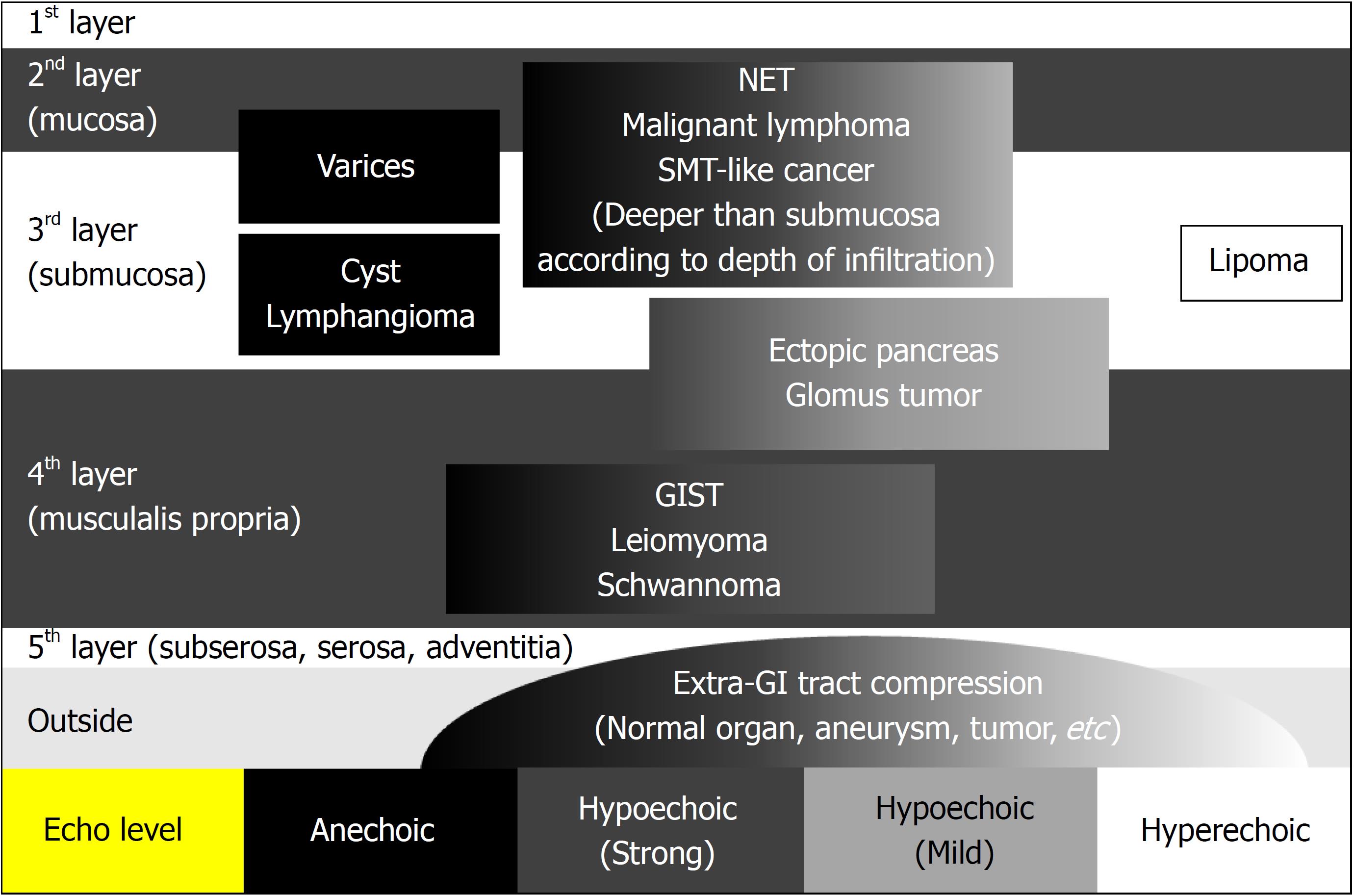
Figure 4 Differential diagnosis of subepithelial lesions by endoscopic ultrasound.
Quoted and modified from reference[39] with permission. GIST: Gastrointestinal stromal tumor.
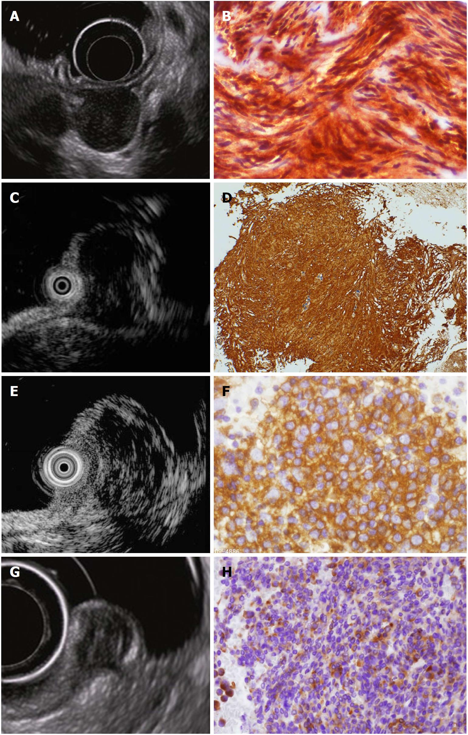
Figure 5 Endoscopic ultrasound images and corresponding endoscopic ultrasonography-guided fine needle aspiration specimens of hypoechoic solid tumors.
A: Endoscopic ultrasound (EUS) image of a gastric gastrointestinal stromal tumor; B: EUS-guided fine needle aspiration (EUS-FNA) specimen tissue image of A (KIT-positive spindle-shaped tumor cells are observed); C: EUS image of gastric leiomyoma; D: EUS-FNA specimen tissue image of C [α-SMA-positive spindle-shaped tumor cells are observed; diagnosis of leiomyoma was made by immunohistochemical analysis, which revealed α-SMA (+), KIT (-), CD34 (-), and S-100 (-)]; E: EUS image of gastric malignant lymphoma; F: EUS-FNA specimen image of E (diagnosis of diffuse large B-cell lymphoma was made by CD20-positive lymphoid tumor cells); G: EUS image of rectal neuroendocrine tumor (NET); H: EUS-FNA specimen image of G (diagnosis of NET was made by typical findings of irregular nest of synaptophysin-positive epithelial-like cells). Quoted and modified from reference[38] with permission.
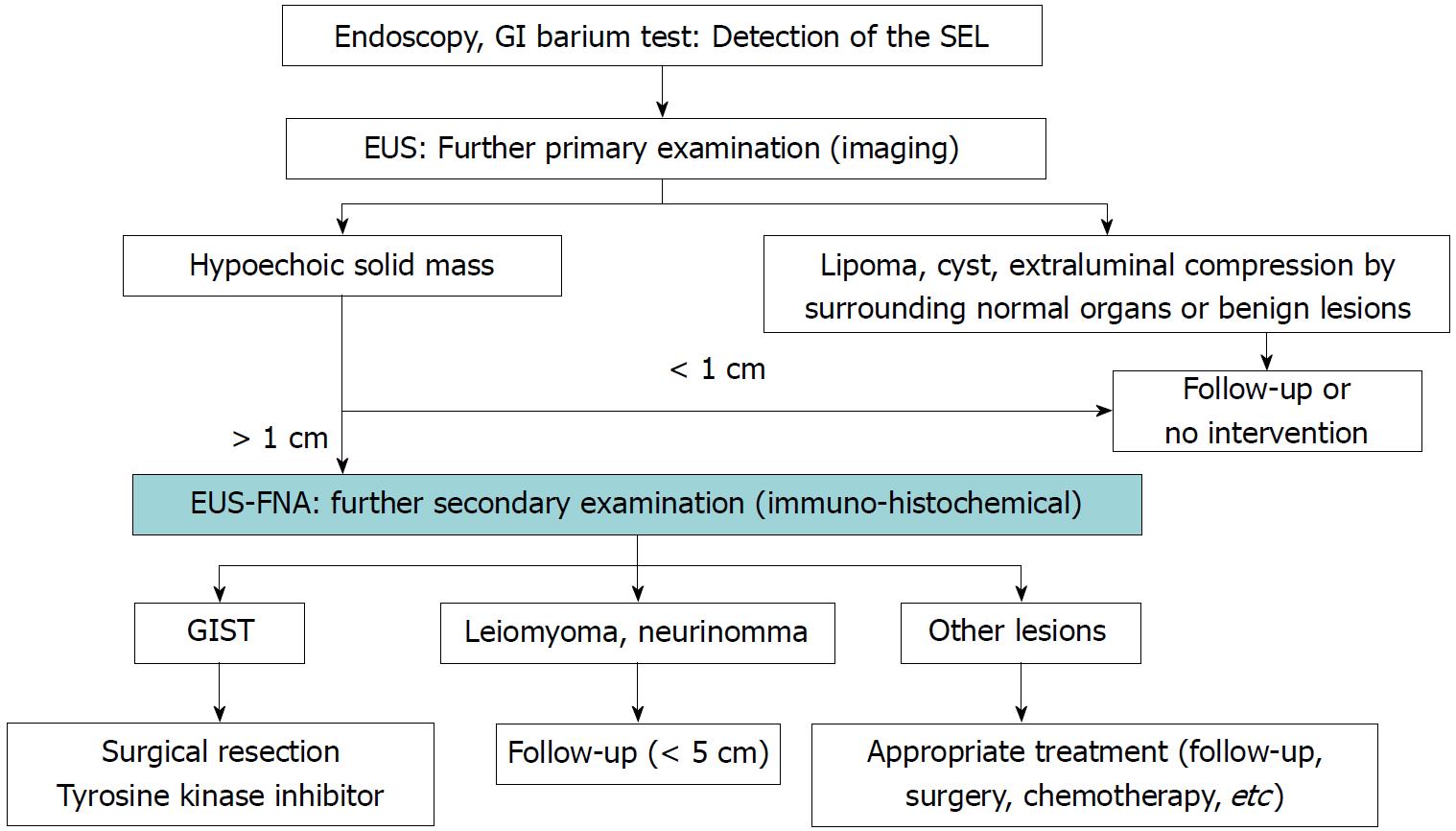
Figure 6 Proposed algorithm for management of subepithelial lesions.
Quoted and modified from reference[21]. GIST: Gastrointestinal stromal tumor; GI: Gastrointestinal; SEL: Subepithelial lesion; EUS: Endoscopic ultrasound; EUS-FNA: Endoscopic ultrasound-guided fine needle aspiration.
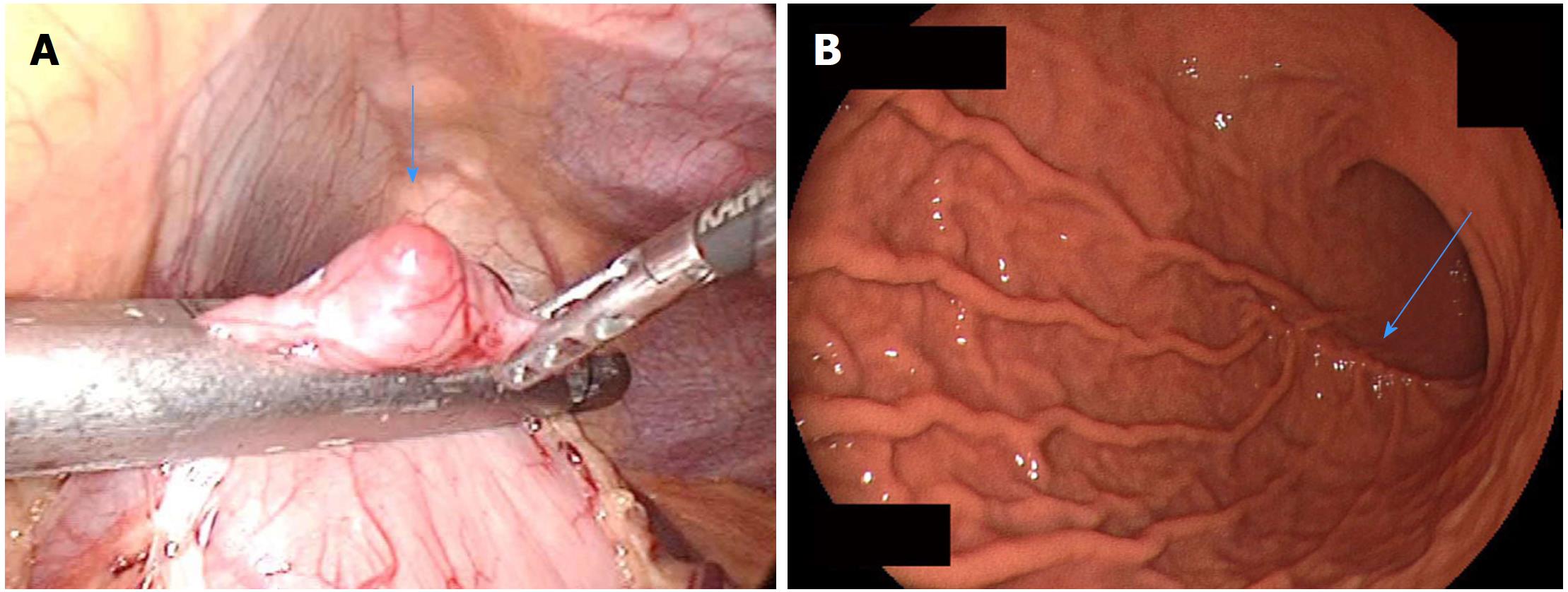
Figure 7 Laparoscopic resection of a small gastric gastrointestinal stromal tumor.
A: Laparoscopic view of a small gastric gastrointestinal stromal tumor (arrow) during resection; B: Postoperative endoscopy shows mild postoperative deformity (arrow). Quoted and modified from reference[77] with permission.
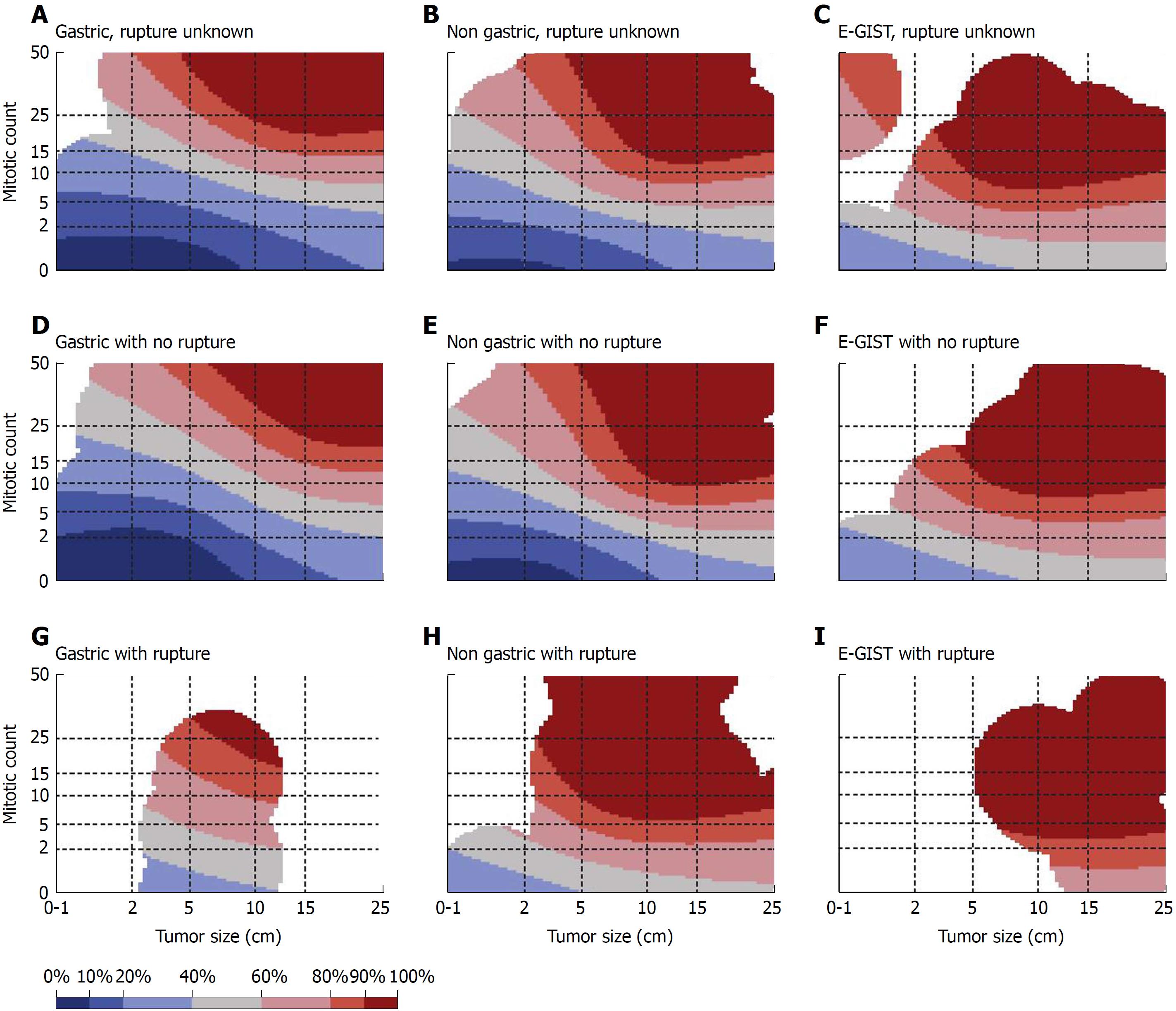
Figure 8 Contour maps for estimating the risk of gastrointestinal stromal tumor recurrence after surgery.
Areas of colors according to the recurrence rate at the 10th year after surgical treatment of GIST: Blue-black: 0%-10%, Blue: 10%-20%, Light blue: 20%-40%, Gray: 40%-60%, Pink: 60%-80%, Red: 80%-90%, Dark red: 90%-100%. Reprinted from reference[87] with permission. E-GIST: Extra-gastrointestinal stromal tumor (arising outside the gastrointestinal tract).
- Citation: Akahoshi K, Oya M, Koga T, Shiratsuchi Y. Current clinical management of gastrointestinal stromal tumor. World J Gastroenterol 2018; 24(26): 2806-2817
- URL: https://www.wjgnet.com/1007-9327/full/v24/i26/2806.htm
- DOI: https://dx.doi.org/10.3748/wjg.v24.i26.2806


