Copyright
©The Author(s) 2018.
World J Gastroenterol. Jul 14, 2018; 24(26): 2806-2817
Published online Jul 14, 2018. doi: 10.3748/wjg.v24.i26.2806
Published online Jul 14, 2018. doi: 10.3748/wjg.v24.i26.2806
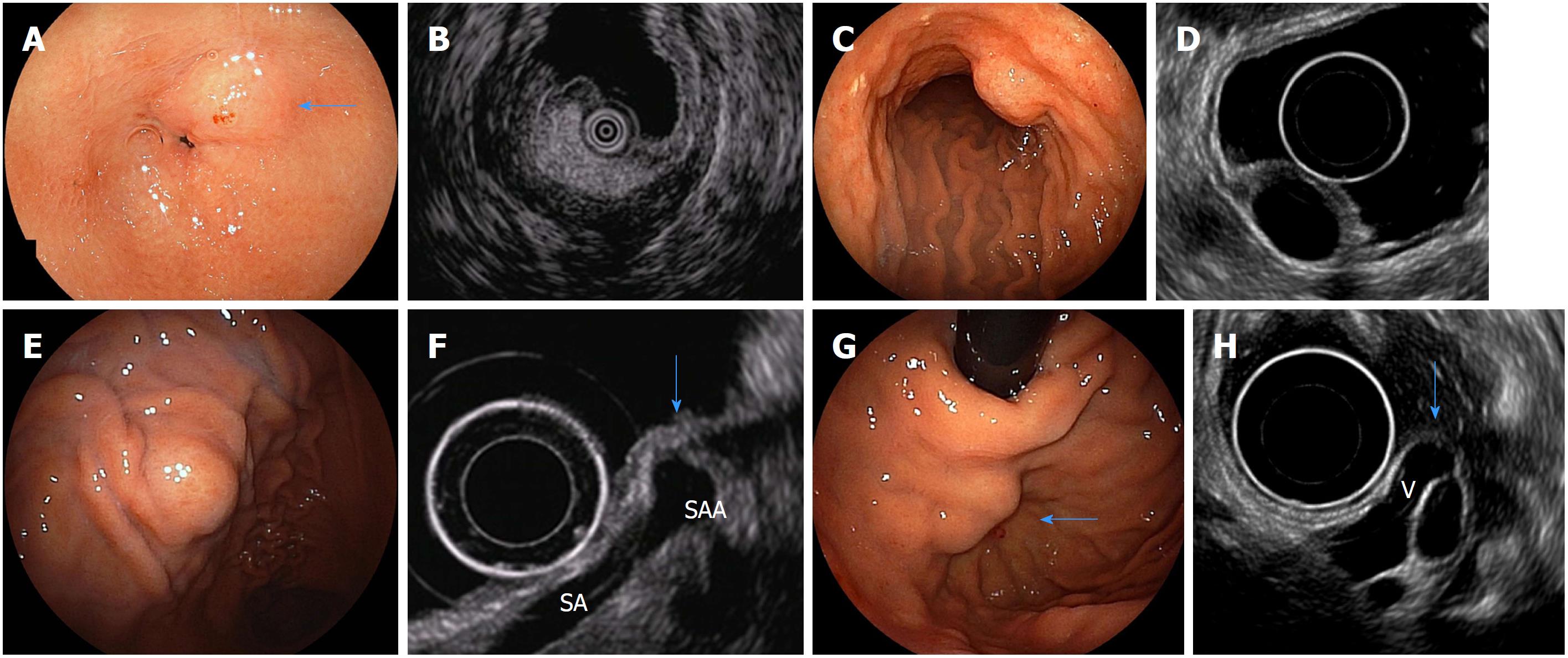
Figure 3 Endoscopic images of subepithelial lesions that can be diagnosed only with endoscopic ultrasound findings and their specific endoscopic ultrasonography images.
A: Endoscopic image of a gastric lipoma (arrow); B: Endoscopic ultrasound (EUS) image of A (high-echo mass); C: Endoscopic image of a gastric cyst; D: EUS image of C (anechoic mass); E: Endoscopic image of extra-gastric compression due to splenic artery aneurysm; F: EUS image of E [normal gastric wall is compressed by a splenic artery aneurysm(SAA) (arrow). SA: splenic artery]; G: Endoscopic image of gastric varices (arrow); H: EUS image of G [varices are present in the submucosa from the outside of the wall (V) (arrow)]. Quoted and modified from reference[38] with permission.
- Citation: Akahoshi K, Oya M, Koga T, Shiratsuchi Y. Current clinical management of gastrointestinal stromal tumor. World J Gastroenterol 2018; 24(26): 2806-2817
- URL: https://www.wjgnet.com/1007-9327/full/v24/i26/2806.htm
- DOI: https://dx.doi.org/10.3748/wjg.v24.i26.2806


