Copyright
©The Author(s) 2018.
World J Gastroenterol. Mar 14, 2018; 24(10): 1120-1133
Published online Mar 14, 2018. doi: 10.3748/wjg.v24.i10.1120
Published online Mar 14, 2018. doi: 10.3748/wjg.v24.i10.1120
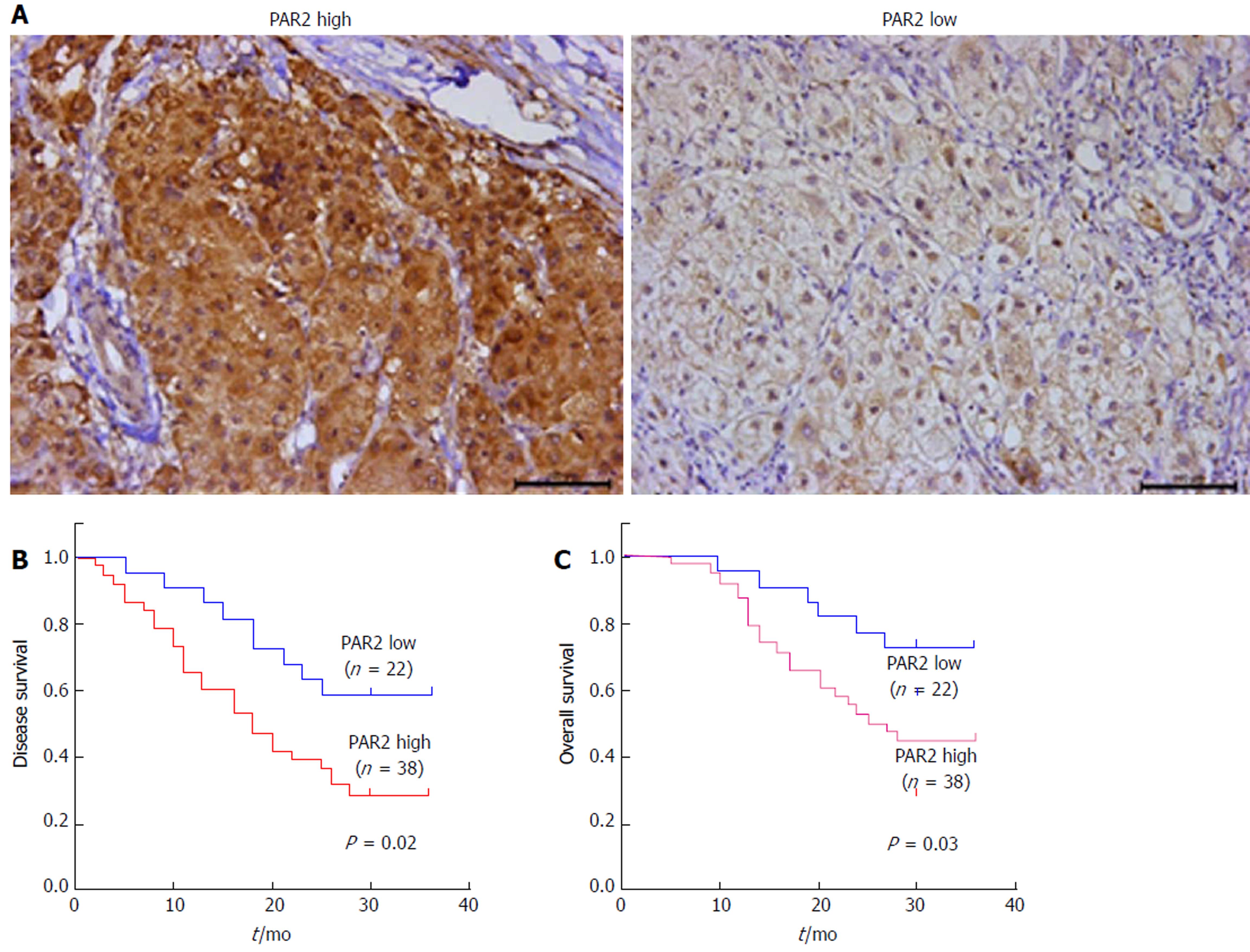
Figure 1 The expression level of proteinase-activated receptor 2 predicts overall survival and disease-free survival in hepatocellular carcinoma patients.
A: Immunohistochemical staining for proteinase-activated receptor 2 (PAR2) in hepatocellular carcinoma (HCC) tissues, and the expression level of PAR2 was divided into high (left) and low (right); B: Disease-free survival in HCC patients correlated with PAR2 expression; C: Overall survival in HCC patients correlated with PAR2 expression.
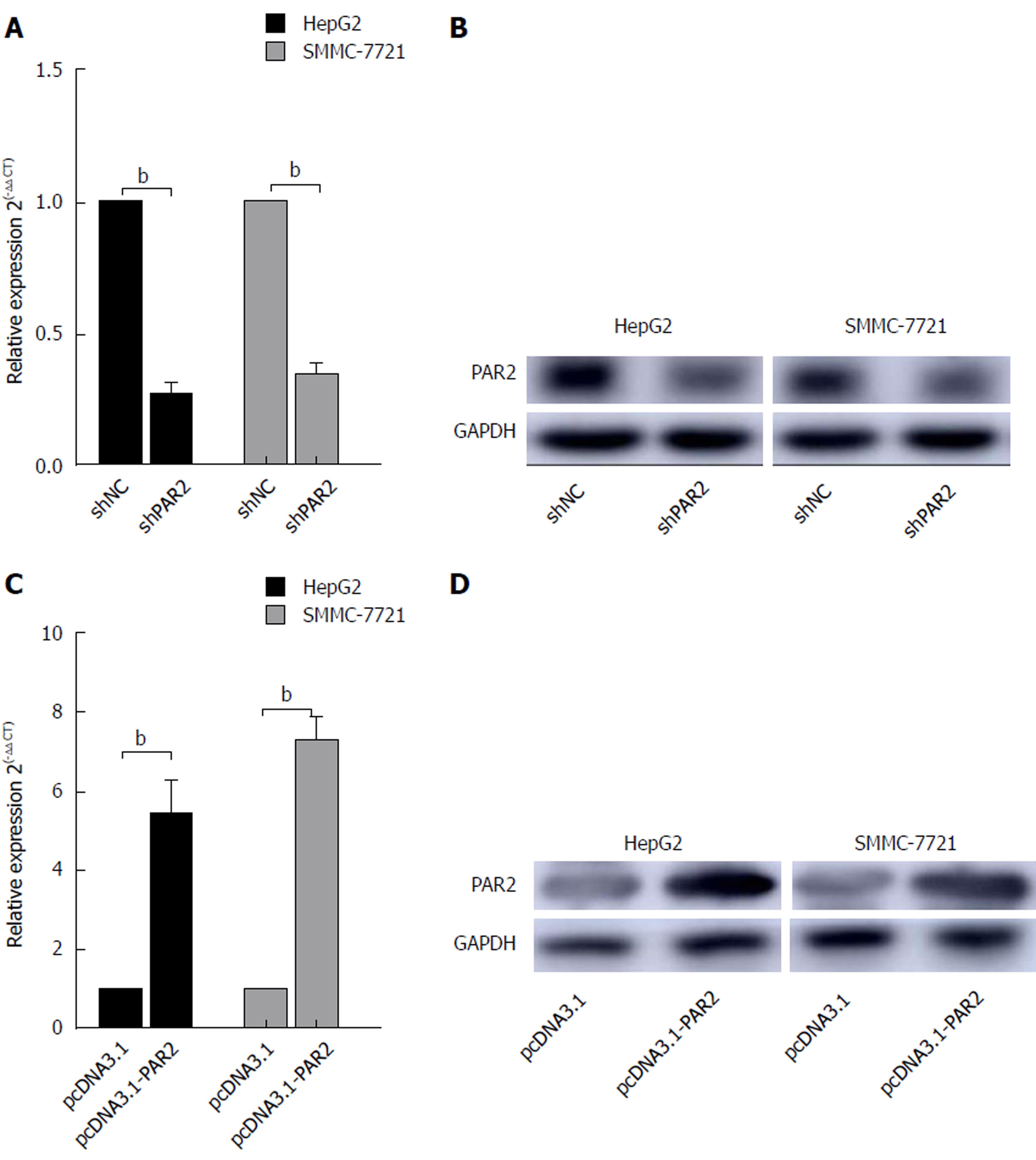
Figure 2 Proteinase-activated receptor 2 knockdown and overexpression.
A: qRT-PCR analysis of the proteinase-activated receptor 2 (PAR2) mRNA expression levels in HepG2 and SMMC-7721 cells with PAR2 knockdown; B: Immunoblot analysis of the protein levels of PAR2 in HepG2 and SMMC-7721 cells with PAR2 knockdown; C: The RNA expression levels of PAR2 in HepG2 and SMMC-7721 cells with PAR2 overexpression were measured by qRT-PCR; D: The protein levels of PAR2 in HepG2 and SMMC-7721 cells with PAR2 overexpression were measured by immunoblot analysis. bP < 0.01.
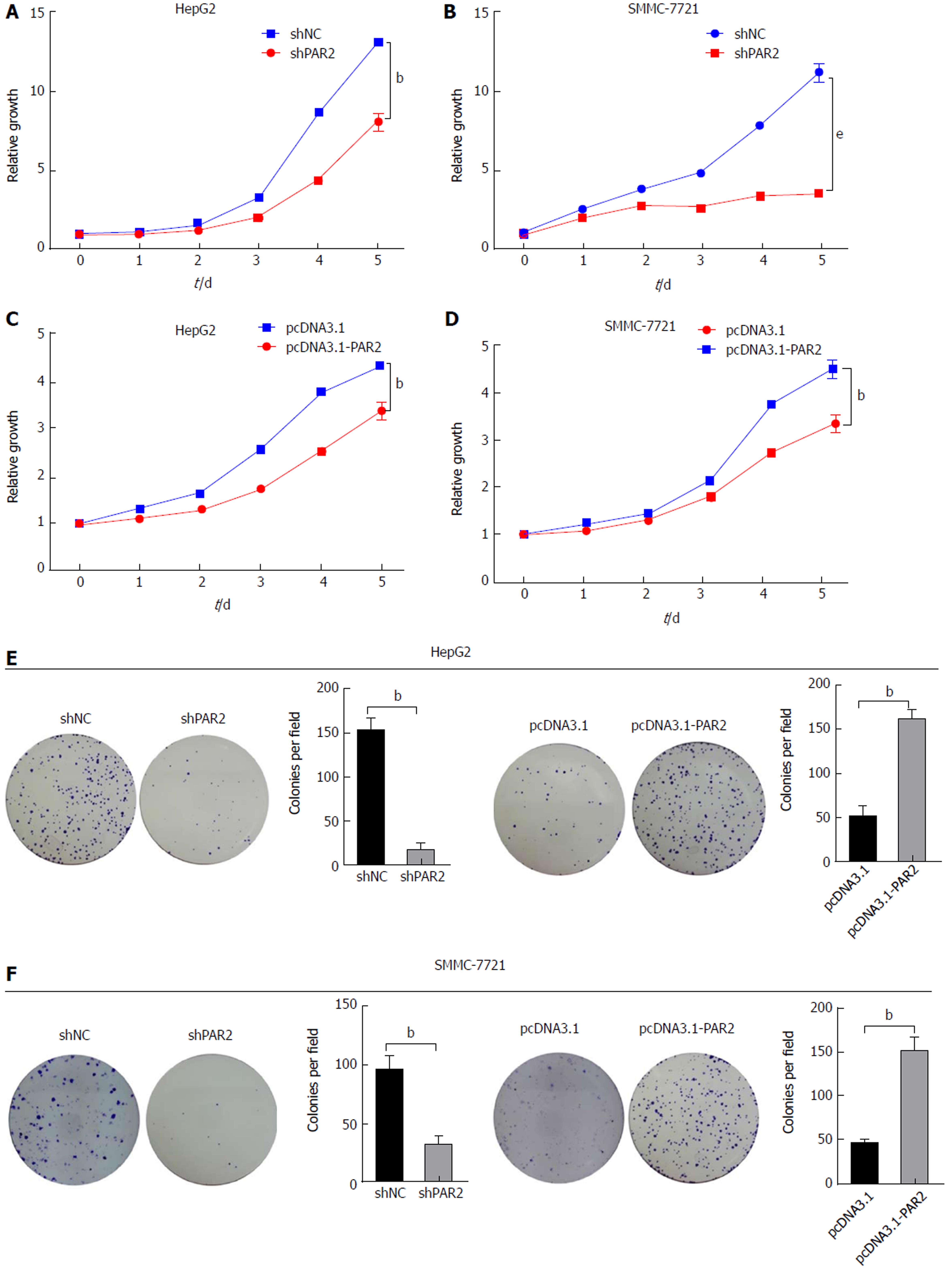
Figure 3 Proteinase-activated receptor 2 promotes proliferation of hepatocellular carcinoma cells.
A-D: CCK8 assay in HepG2 and SMMC-7721 cells; E and F: Colony formation assay in HepG2 and SMMC-7721 cells. The colony number in each field was counted. bP < 0.01, eP < 0.001.
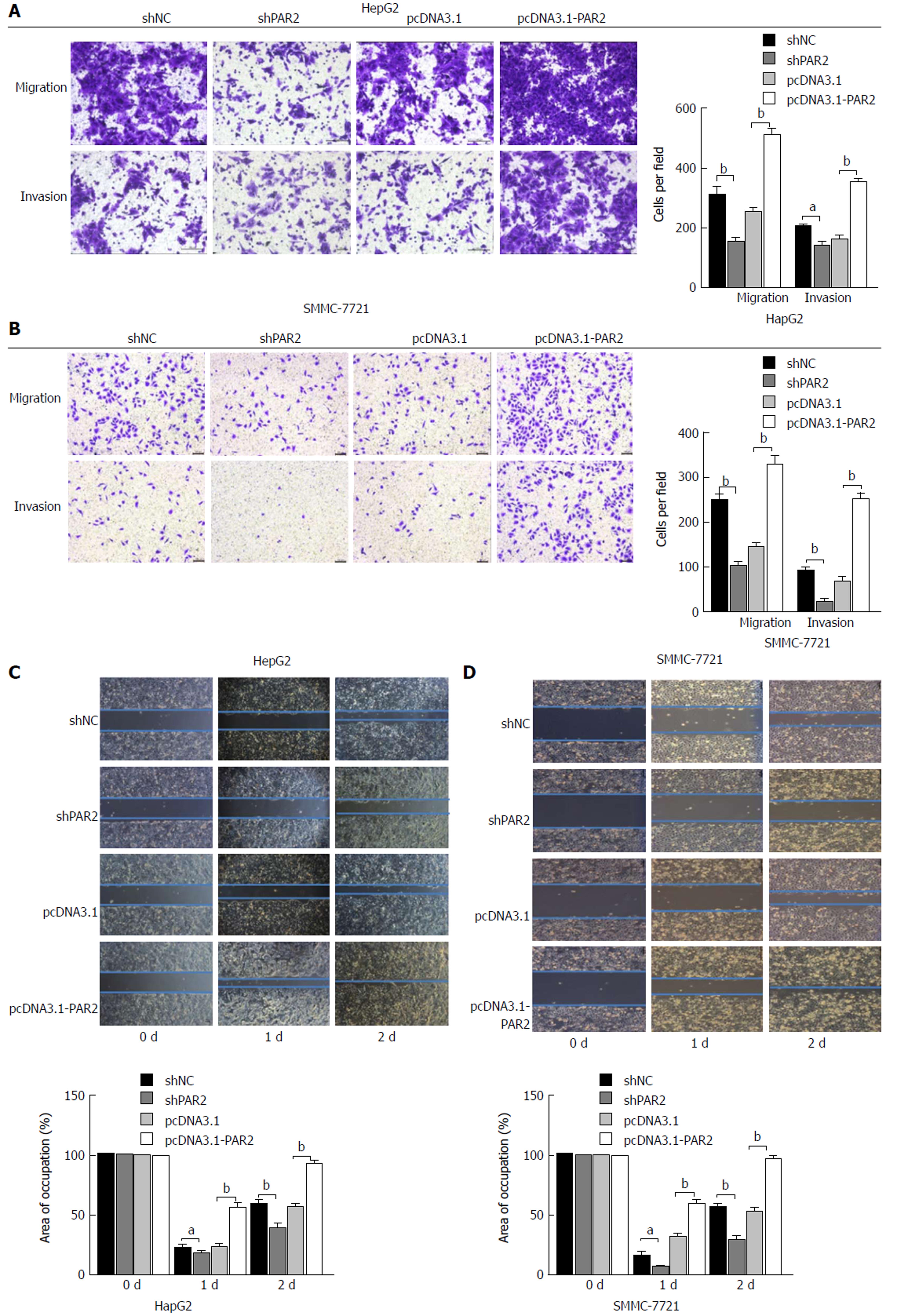
Figure 4 Proteinase-activated receptor 2 promotes hepatocellular carcinoma cell invasion and migration in vitro.
A and B: Transwell assays with or without matrigel coating were performed in HepG2 and SMMC-7721 cells. Cell number in each field was counted; C and D: Wound healing assays were performed in HepG2 and SMMC-7721 cells and the pictures were taken every 24 h (aP < 0.05, bP < 0.01).
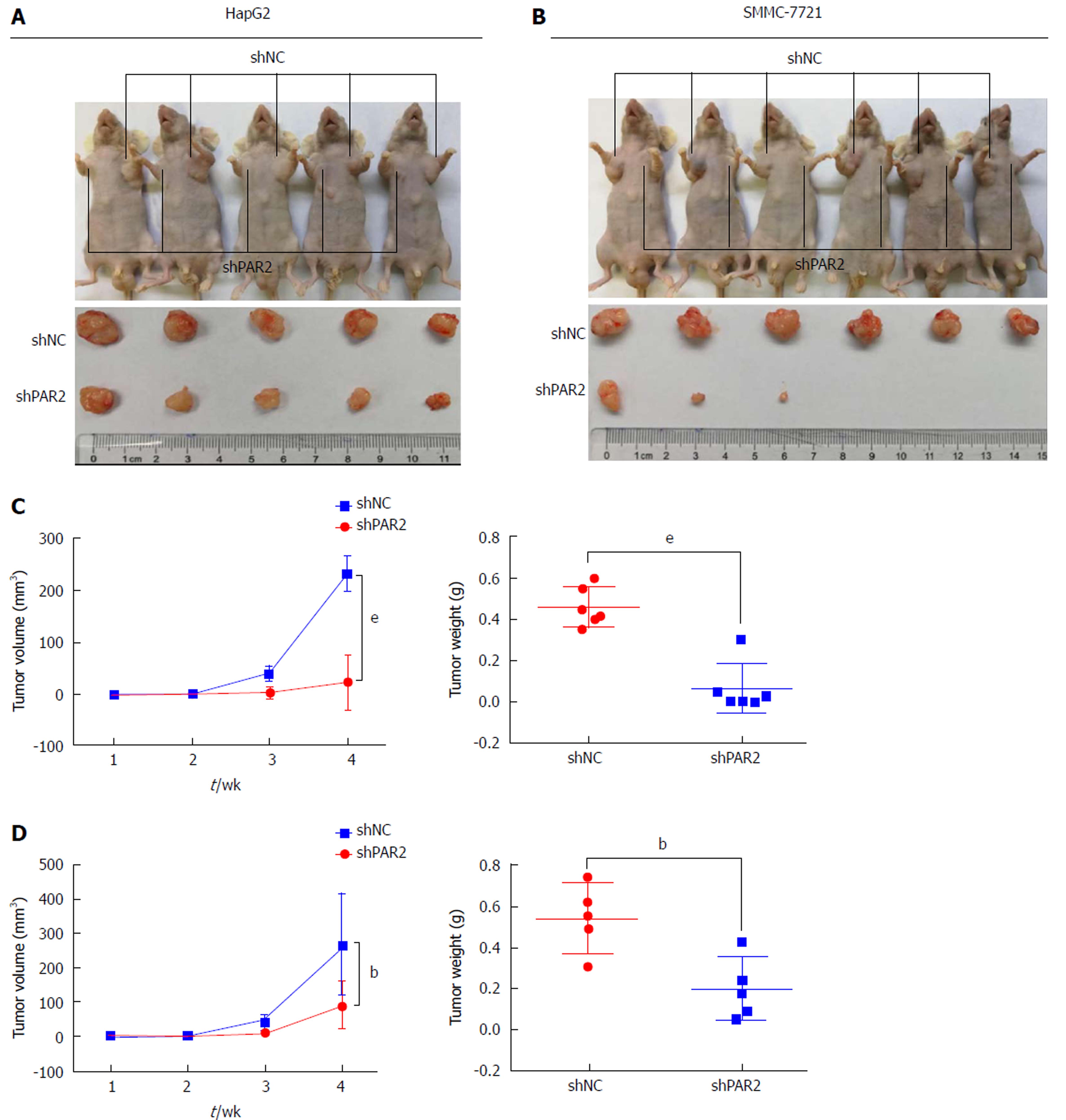
Figure 5 Proteinase-activated receptor 2 knockdown inhibits tumor growth in nude mice.
A and B: Subcutaneous xenografts of HepG2 and SMMC-7721 cells in nude mice growing for 4 wk; C and D: Tumor volumes were measured every week and tumor weights were tested at the end. bP < 0.01, eP < 0.001.
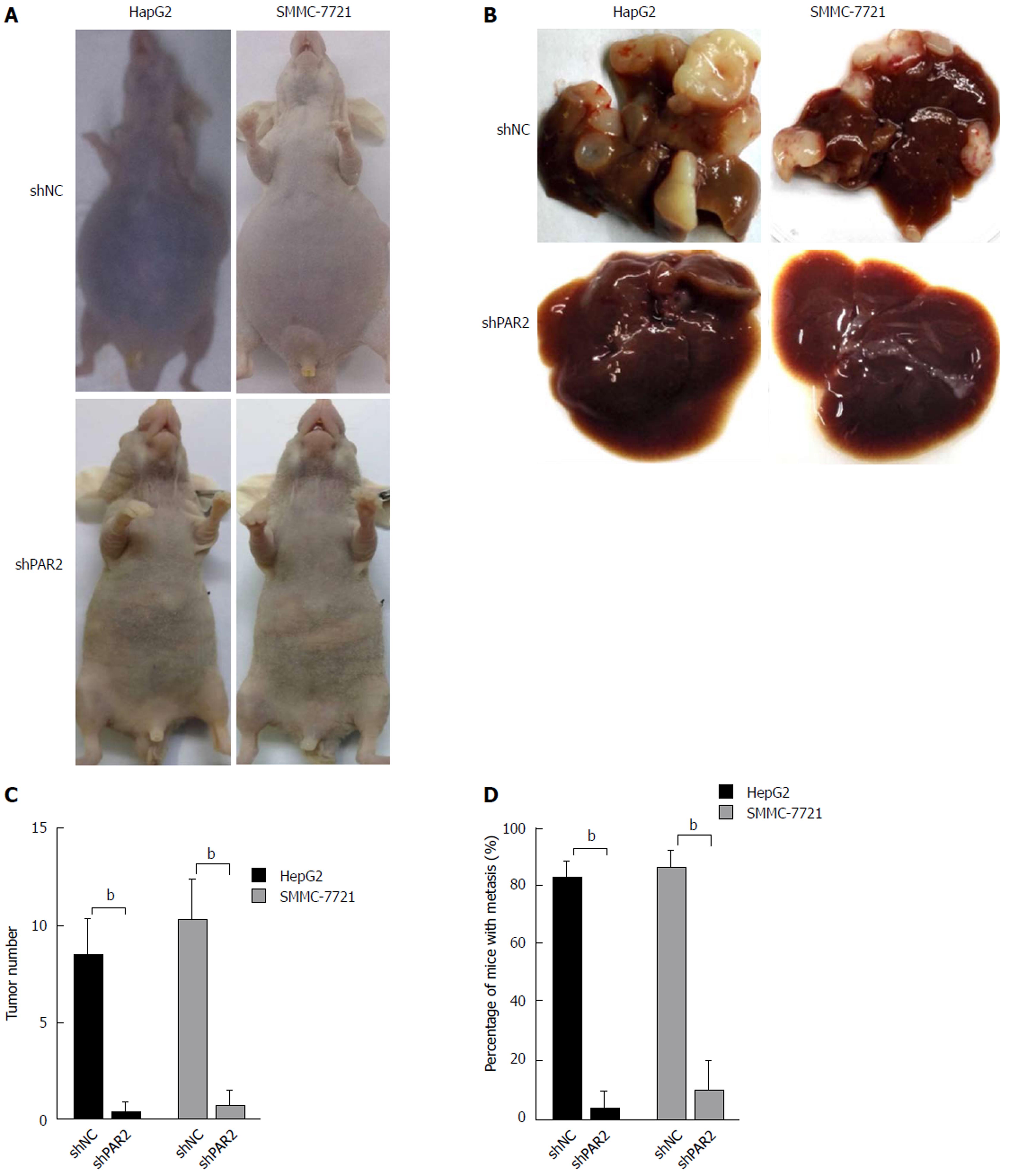
Figure 6 Proteinase-activated receptor 2 knockdown inhibits tumor metastasis in nude mice.
A: HepG2 and SMMC-7721 cells were injected into the spleen of nude mice to form liver metastases. The appearance of mice after 4 wk is shown; B: Tumors grown on the liver; C: Tumor number on each liver was counted; D: Percentage of mice with liver metastases (n = 10 per group). bP < 0.01.
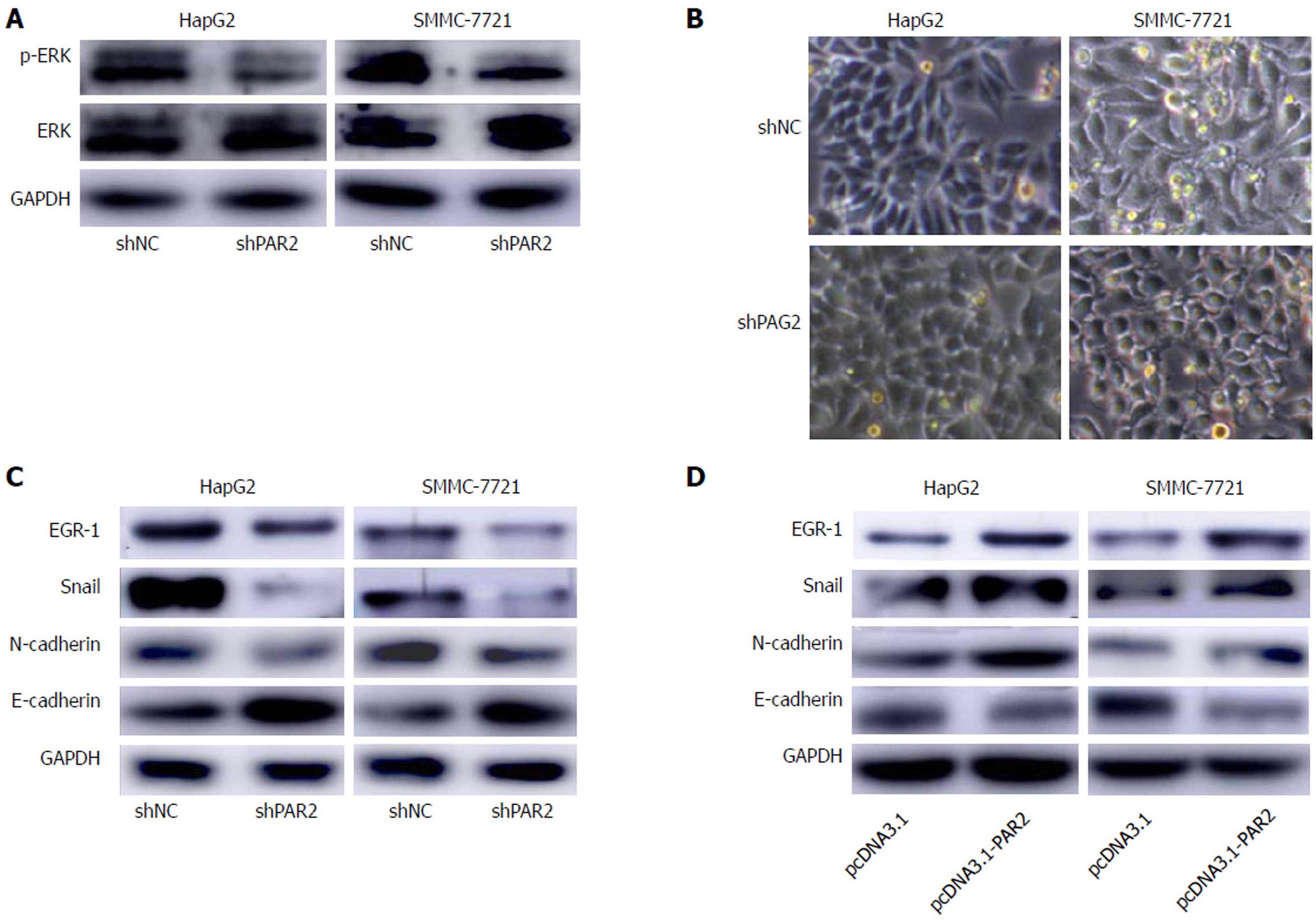
Figure 7 Proteinase-activated receptor 2 mediates the activation of ERK and further induces epithelial-mesenchymal transition.
A: The protein levels of ERK and p-ERK in HepG2 and SMMC-7721 cells; B: The morphological changes in HepG2 and SMMC-7721 cells after proteinase-activated receptor 2 knockdown; C and D: The protein levels of EGR-1, Snail, E-cadherin, and N-cadherin in HepG2 and SMMC-7721 cells. GAPDH was selected as an endogenous control.
- Citation: Sun L, Li PB, Yao YF, Xiu AY, Peng Z, Bai YH, Gao YJ. Proteinase-activated receptor 2 promotes tumor cell proliferation and metastasis by inducing epithelial-mesenchymal transition and predicts poor prognosis in hepatocellular carcinoma. World J Gastroenterol 2018; 24(10): 1120-1133
- URL: https://www.wjgnet.com/1007-9327/full/v24/i10/1120.htm
- DOI: https://dx.doi.org/10.3748/wjg.v24.i10.1120


