Copyright
©The Author(s) 2016.
World J Gastroenterol. Jun 21, 2016; 22(23): 5364-5373
Published online Jun 21, 2016. doi: 10.3748/wjg.v22.i23.5364
Published online Jun 21, 2016. doi: 10.3748/wjg.v22.i23.5364
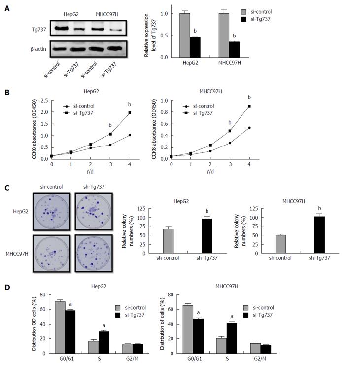
Figure 1 Down-regulation of Tg737 accelerates hepatocellular carcinoma cell proliferation.
A: Western blot analysis of Tg737 expression in HepG2 and MHCC97H cells transfected with si-Tg737 or negative control (si-control); B: Impact of Tg737 down-regulation on HCC cell proliferation detected by CCK-8 assay after si-Tg737 and si-control transfection in HepG2 and MHCC97H cell lines; C: Impact of Tg737 down-regulation on colony forming ability of HepG2 and MHCC97H cell lines. The relative percentage of colony numbers from the si-Tg737 group is given as 100%; D: Cell cycle assay of HepG2 and MHCC97H cells transfected with si-Tg737 and si-control. The charts were illustrated by the percentage of cells in the G0/G1, S, and G2/M phase of cell cycle, respectively. aP < 0.05, bP < 0.01 vs control.
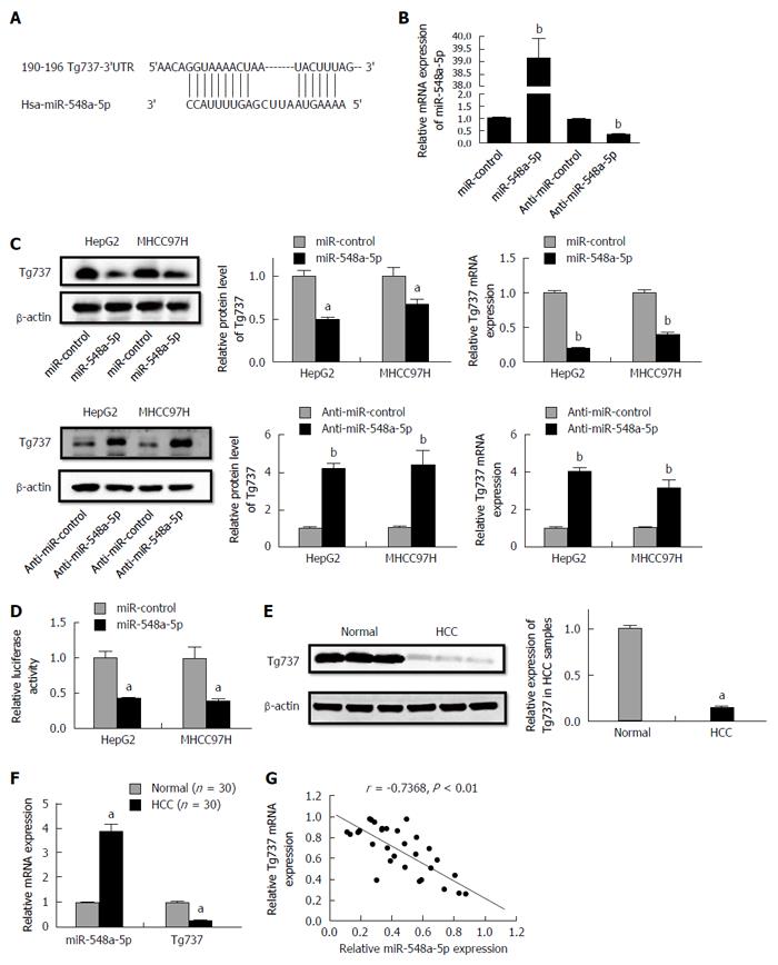
Figure 2 Tg737 is a target gene of miR-548a-5p.
A: MiR-548a-5p and its deductive binding domain in the 3'UTR of Tg737; B: Relative expression of miR-548a-5p; C: qRT-PCR and Western blot analysis demonstrated that miR-548a-5p overexpression significantly decreased Tg737 mRNA expression and miR-548a-5p knockdown significantly increased Tg737 mRNA expression in HepG2 and MHCC97H cells; D: Luciferase reporter assay of the connection between the 3'UTR of Tg737 and miR-548a-5p in HepG2 and MHCC97H cells; E: Western blot analysis demonstrated higher miR-548a-5p and lower Tg737 expression in human HCC specimens compared to normal liver tissue; F: qRT-PCR demonstrated higher miR-548a-5p and lower Tg737 expression in human HCC specimens compared to normal liver tissue; G: Spearman’s correlation assay showed that miR-548a-5p was negatively correlated with Tg737 mRNA expression in HCC specimens. aP < 0.05, bP < 0.01 vs control.
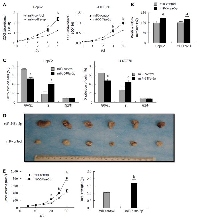
Figure 3 miR-548a-5p promotes hepatocellular carcinoma cell proliferation in vitro and in vivo.
A: CCK-8 assays of HepG2 and MHCC97H cells constantly expressing miR-548a-5p or miR-control; B: Colony forming assays of HepG2 and MHCC97H cells constantly expressing miR-548a-5p or miR-control; C: Cell cycle assays of HepG2 and MHCC97H cells constantly expressing miR-548a-5p or miR-control; D: MiR-548a-5p or miR-control overexpressing HepG2 cells were injected into the subcutaneous layer of nude mice. The mice were killed 30 d after injection; E: Tumor volumes and weight. aP < 0.05, bP < 0.01 vs control.
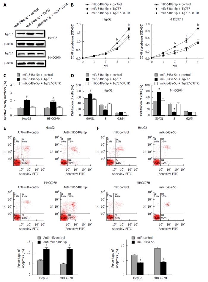
Figure 4 Restoration of Tg737 rescues the miR-548a-5p induced cell proliferation inhibition and apoptosis induced by cisplatin.
A: Western blot was performed 48 h after transfection of pc CON, pc Tg737 and plasmid with 3’UTR of Tg737 into miR-548a-5p overexpressing HepG2 and MHCC97H cells; B: CCK-8 assay; C: Colony forming assay; D: Cell cycle assay; E: Cell apoptosis increased in anti-miR-548a-5p transfected HepG2 and MHCC97H cells; F: Cell apoptosis decreased in miR-548a-5p transfected HepG2 and MHCC97H cells. aP < 0.05, bP < 0.01 vs control.
- Citation: Zhao G, Wang T, Huang QK, Pu M, Sun W, Zhang ZC, Ling R, Tao KS. MicroRNA-548a-5p promotes proliferation and inhibits apoptosis in hepatocellular carcinoma cells by targeting Tg737. World J Gastroenterol 2016; 22(23): 5364-5373
- URL: https://www.wjgnet.com/1007-9327/full/v22/i23/5364.htm
- DOI: https://dx.doi.org/10.3748/wjg.v22.i23.5364


