Copyright
©The Author(s) 2016.
World J Gastroenterol. Jun 7, 2016; 22(21): 4999-5011
Published online Jun 7, 2016. doi: 10.3748/wjg.v22.i21.4999
Published online Jun 7, 2016. doi: 10.3748/wjg.v22.i21.4999
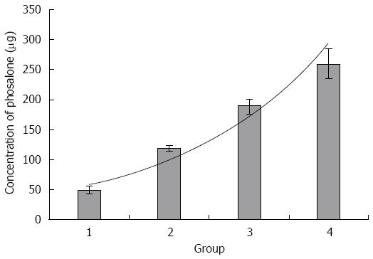
Figure 1 Determination of LD50 of phosalone.
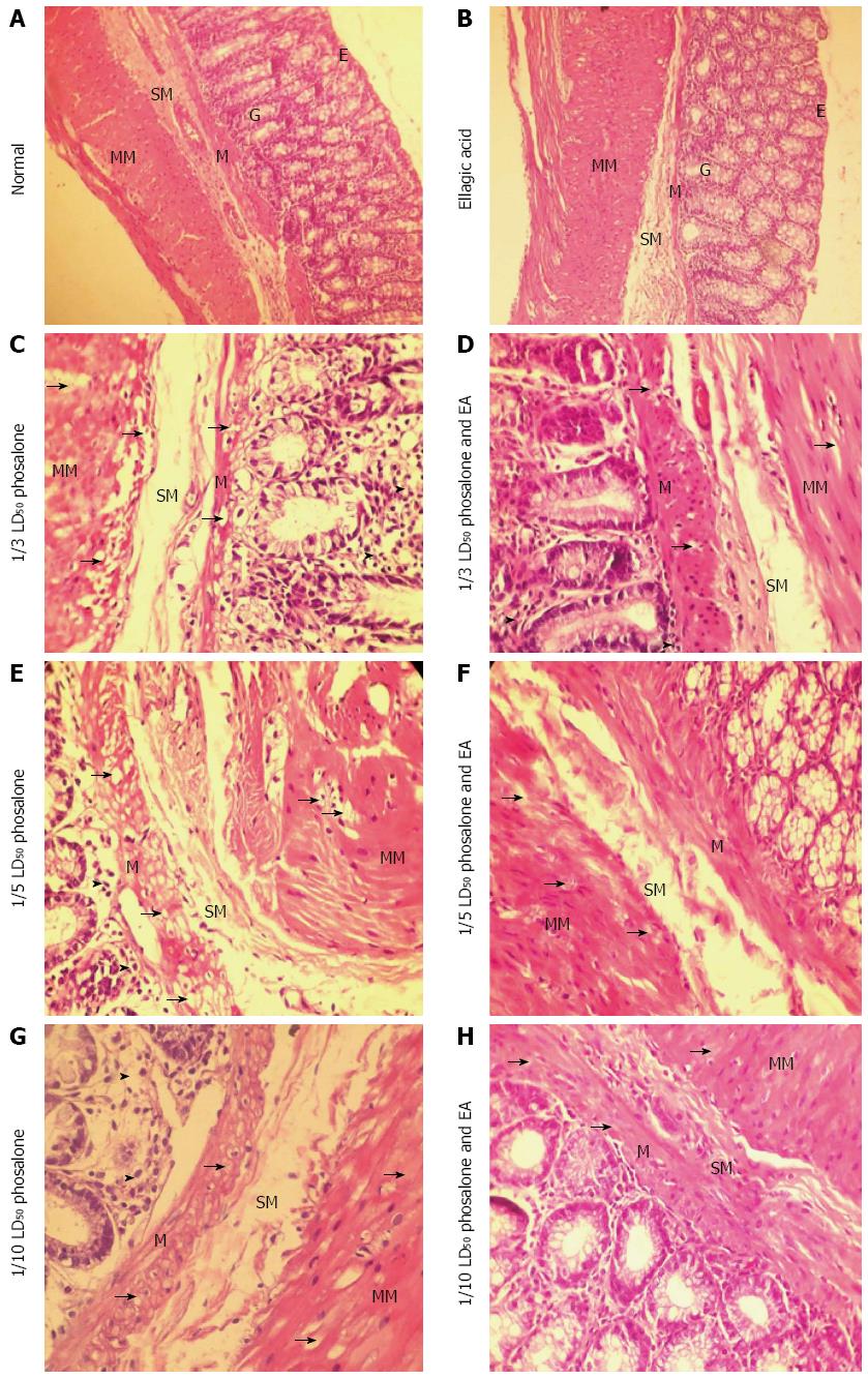
Figure 2 Histological images of colon tissues from normal, ellagic acid and experimental groups.
In control group, different parts of the colon tissue are healthy. There are no erosions or ulcers in epithelium. It cannot be seen any necroses and inflammation cells in mucus and mucosal glands in the lamina propria. The mucus thickness, the size of the glands, the muscle layer of mucosal and serous is normal. No degeneration, swelling and goblet cells are observable (A). In ellagic acid (EA) group, the following are normal, the mucosal, the goblet gland cells, epithelium, the mucosa thickness and the size of the glands. There are no ulcers, necrosis hyperplasia and inflammation cells such as neutrophils or lymphocytes. It cannot be observed any degeneration, swelling and goblet cells. The serous is normal without any adherent (B). In 1/3 LD50 phosalone group, no necrosis and ulcer are visible in epithelium. The mucosal glands are normal but severe degeneration of mucosal muscle cells and muscle layers is observable. In addition to hyperemia, there is a significant infiltration of mononuclear inflammatory cells such as lymphocytes and plasma cells between the mucosal glands. There is no fibrosis and serous adhesion (C). In 1/3 LD50 phosalone group and EA, no necrosis and ulcer are visible in epithelium. The mucosal glands are normal but mild degeneration of mucosal muscle cells and muscle layers is observable. The level of degeneration and inflammation is less than 1/3 LD50 phosalone group. There is a very mild inflammation due to lymphocytes infiltration between mucosal glands. There is no serous adhesion (D). In 1/5 LD50 phosalone group, no necrosis and ulcer are visible in epithelium. The mucosal glands are normal but relatively severe degeneration of mucosal muscle cells and muscle layers is observable. In addition to hyperemia, there is a significant infiltration of mononuclear inflammatory cells such as lymphocytes and plasma cells between the mucosal glands. There is no fibrosis and serous adhesion (E). In 1/5 LD50 phosalone group and EA, no necrosis and ulcer are visible in epithelium. The mucosal glands are normal but mild degeneration of mucosal muscle cells and muscle layers is observable. The level of degeneration is less than 1/5 LD50 phosalone group. There is a very mild inflammation due to lymphocytes infiltration between mucosal glands. There is no serous adhesion (F). In 1/10 LD50 phosalone group, no necrosis and ulcer is present in epithelial. The mucosal glands are normal but significant degeneration of muscle cells and mucosal layer is observable. In addition to hyperemia, there is a mild infiltration of mononuclear inflammatory cells such as lymphocytes between the mucosal glands. There is no fibrosis and serous adhesion (G). In 1/10 LD50 phosalone group and EA, no necrosis and ulcer are visible in epithelium. The mucosal glands are normal but mild degeneration of mucosal muscle cells and muscle layers is observable. The level of degeneration is less than 1/10 LD50 phosalone group. There is a very mild inflammation due to lymphocytes infiltration between mucosal glands. There is no serous adhesion (H). In 1/20 LD50 phosalone group, there is no evidence of necroses, ulcer or inflammation in epithelium. Mucosal glands are normal but a mild degeneration is observable in muscle cells in mucosal layer. A mild diapedesis, hyperemia and inflammation in mucosal is visible. There is no fibrosis and serous adhesion (I). In 1/20 LD50 phosalone group and EA, no necrosis and ulcer are visible in epithelium. The mucosal glands are normal but mild degeneration of mucosal muscle cells and muscle layers is observable. The level of degeneration is less than 1/20 LD50 phosalone group. There is no inflammation in different layers (J). ML: Muscular layer; SM: SubMucosa; M: Mucosa; G: Gland; E: Epithelium.
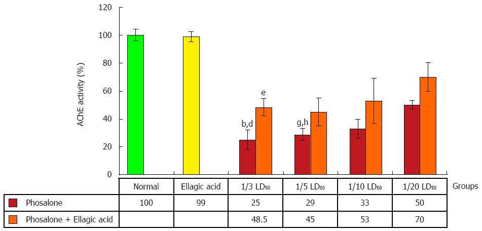
Figure 3 Effect of phosalone and ellagic acid on AChE activity of colon cells.
Values are mean ± SE. bP < 0.001 vs normal group; dP < 0.001 vs ellagic acid group; Ellagic acid significantly increased of AChE activity in 1/3 dose of phosalone group. eP < 0.05 vs (1/3 LD50 phosalone) group; gP < 0.05 vs ellagic acid group, hP < 0.01 vs normal group.
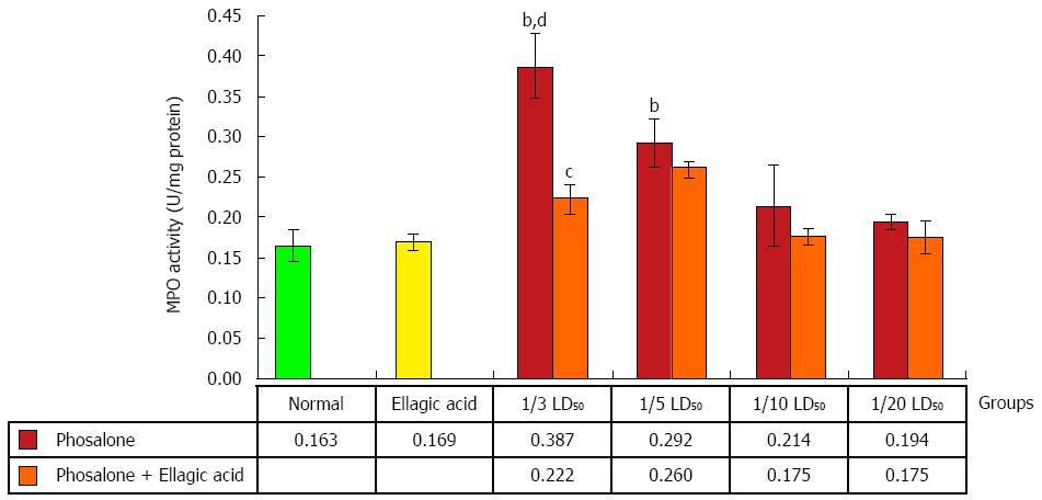
Figure 4 Effect of phosalone and ellagic acid on myeloperoxidase activity of colon cells.
Ellagic acid significantly decreased of MPO in 1/3 dose of phosalone group. Values are mean ± SE. bP < 0.001 vs normal group; cP < 0.05 vs (1/3 LD50 phosalone) group; dP < 0.01 vs Ellagic acid group. MPO: Myeloperoxidase activity.
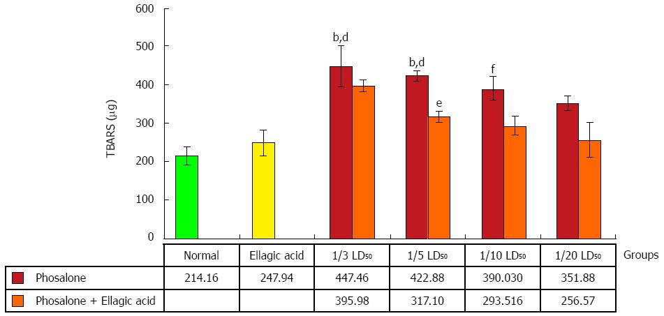
Figure 5 Effect of phosalone and ellagic acid on oxidative-stress as thiobarbituric acid-reaction substances of colon cells.
Ellagic acid significantly decreased of thiobarbituric acid-reaction substances in 1/5 dose of phosalone group. Values are mean ± SE. bP < 0.001 vs normal group; dP < 0.01 vs Ellagic acid group; eP < 0.05 vs (1/5 LD50 phosalone) group; fP < 0.01 vs normal group.
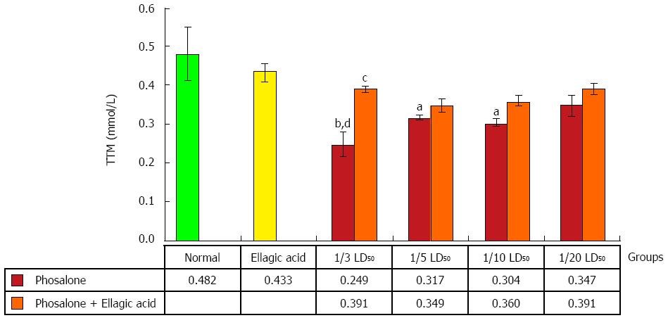
Figure 6 Effect of phosalone and ellagic acid on total thiol molecules activity of colon cells.
Ellagic acid significantly increased of total thiol molecules in 1/3 dose of phosalone group. Values are mean ± SE. aP < 0.05 vs normal group; bP < 0.01 vs normal group; cP < 0.05 vs (1/3 LD50 phosalone) group; dP < 0.01 vs Ellagic acid group.
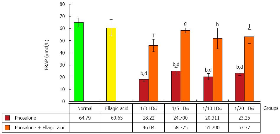
Figure 7 Effect of phosalone and ellagic acid on anti-oxidant power as (ferric reducing antioxidant power) of colon cells.
Ellagic acid significantly decreased of ferric reducing antioxidant power in all doses of phosalone groups. Values are mean ± SE. bP < 0.001, vs normal group; dP < 0.001 vs ellagic acid group; fP < 0.01 vs (1/3 LD50 phosalone) group; gP < 0.001 vs 1/5 LD50 phosalone; hP < 0.001 vs 1/10 LD50 phosalone; jP < 0.01 vs 1/20 LD50 phosalone.
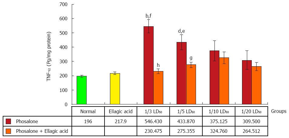
Figure 8 Effect of phosalone and ellagic acid on tumor necrosis factor-α of colon cells.
Ellagic acid significantly decreased of tumor necrosis factor-α in 1/3 and 1/5 doses of phosalone groups. Values are mean ± SE. bP < 0.001 vs normal group; dP < 0.01 vs normal group; eP < 0.05 vs ellagic acid group; fP < 0.01 vs ellagic acid group; gP < 0.05 vs (1/5 LD50 phosalone) group; hP < 0.001 vs (1/3 LD50 phosalone) group.
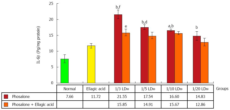
Figure 9 Effect of phosalone and ellagic acid on interlukin-6β of colon cells.
Ellagic acid significantly decreased of tumor necrosis factor-α in 1/3 dose of phosalone group. Values are mean ± SE. aP < 0.05 vs ellagic acid group; bP < 0.001 vs normal group; dP < 0.01 vs ellagic acid group; eP < 0.05 vs (1/3 LD50 phosalone) group; fP < 0.001 vs ellagic acid group.
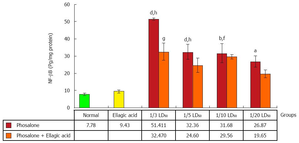
Figure 10 Effect of phosalone and ellagic acid on nuclear factor-κB of colon cells.
Ellagic acid significantly decreased of nuclear factor-κB in 1/3 dose of phosalone group. Values are mean ± SE. aP < 0.05 vs normal group; bP < 0.01 vs normal group; dP < 0.001 vs normal group; fP < 0.01 vs ellagic acid group; hP < 0.001 vs ellagic acid group; Significantly different from at gP < 0.05 vs (1/3 LD50 phosalone) group.
- Citation: Ghasemi-Niri SF, Maqbool F, Baeeri M, Gholami M, Abdollahi M. Phosalone-induced inflammation and oxidative stress in the colon: Evaluation and treatment. World J Gastroenterol 2016; 22(21): 4999-5011
- URL: https://www.wjgnet.com/1007-9327/full/v22/i21/4999.htm
- DOI: https://dx.doi.org/10.3748/wjg.v22.i21.4999


