Copyright
©The Author(s) 2016.
World J Gastroenterol. May 7, 2016; 22(17): 4421-4426
Published online May 7, 2016. doi: 10.3748/wjg.v22.i17.4421
Published online May 7, 2016. doi: 10.3748/wjg.v22.i17.4421
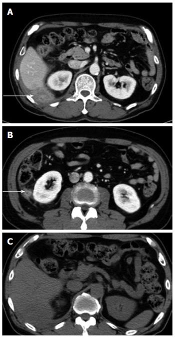
Figure 1 Five months after primary laparoscopic cholecystectomy, computed tomography scan was performed for evaluation of right upper quadrant pain.
A and B: Abdominal CT scan revealed a 4.9 cm × 4.6 cm sized, ill-defined heterogenous retroperitoneal mass in posterolateral sub-hepatic and posterior perirenal space involving posterior abdominal wall (arrows); C: No calcified stone was seen to suggest spilled gallstones in the pre-contrast enhanced CT scan. CT: Computed tomography.
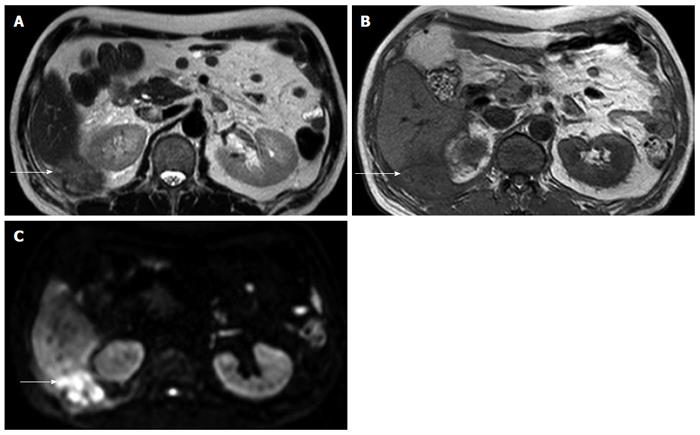
Figure 2 Magnetic resonance imaging showed ill-defined mass at right subhepatic space (arrows).
A: Subtle high signal intensity in T2 weighted image; B: Low signal intensity in T1 weighted image; C: High signal intensity in diffusion weighted image.
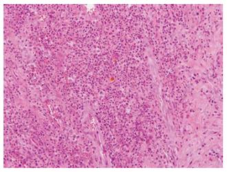
Figure 3 Numerous acute inflammatory cell and limphoplasma cell surrounding fibrosis and granulation tissue is seen.
Bile stained gallstone sludge is seen in the center (haematoxylin and eosin staining, original magnification × 200).
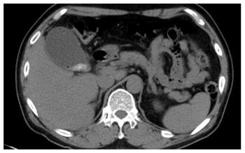
Figure 4 Computed tomography scan showing emphysematous gallbladder and gallbladder stones.
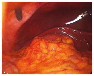
Figure 5 Operative findings in the primary procedure.
Due to edematous, friable wall and gallbladder distension, it was hard to grasp the gallbladder during the traction and the dissection of the gallbladder from the liver.
Figure 6 Multiple spilled gallstones.
Copious irrigation of Morrison’s pouch was applied.
- Citation: Kim BS, Joo SH, Kim HC. Spilled gallstones mimicking a retroperitoneal sarcoma following laparoscopic cholecystectomy. World J Gastroenterol 2016; 22(17): 4421-4426
- URL: https://www.wjgnet.com/1007-9327/full/v22/i17/4421.htm
- DOI: https://dx.doi.org/10.3748/wjg.v22.i17.4421


