Copyright
©The Author(s) 2016.
World J Gastroenterol. May 7, 2016; 22(17): 4421-4426
Published online May 7, 2016. doi: 10.3748/wjg.v22.i17.4421
Published online May 7, 2016. doi: 10.3748/wjg.v22.i17.4421
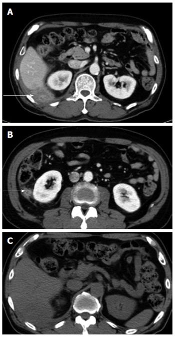
Figure 1 Five months after primary laparoscopic cholecystectomy, computed tomography scan was performed for evaluation of right upper quadrant pain.
A and B: Abdominal CT scan revealed a 4.9 cm × 4.6 cm sized, ill-defined heterogenous retroperitoneal mass in posterolateral sub-hepatic and posterior perirenal space involving posterior abdominal wall (arrows); C: No calcified stone was seen to suggest spilled gallstones in the pre-contrast enhanced CT scan. CT: Computed tomography.
- Citation: Kim BS, Joo SH, Kim HC. Spilled gallstones mimicking a retroperitoneal sarcoma following laparoscopic cholecystectomy. World J Gastroenterol 2016; 22(17): 4421-4426
- URL: https://www.wjgnet.com/1007-9327/full/v22/i17/4421.htm
- DOI: https://dx.doi.org/10.3748/wjg.v22.i17.4421


