Copyright
©The Author(s) 2016.
World J Gastroenterol. May 7, 2016; 22(17): 4421-4426
Published online May 7, 2016. doi: 10.3748/wjg.v22.i17.4421
Published online May 7, 2016. doi: 10.3748/wjg.v22.i17.4421
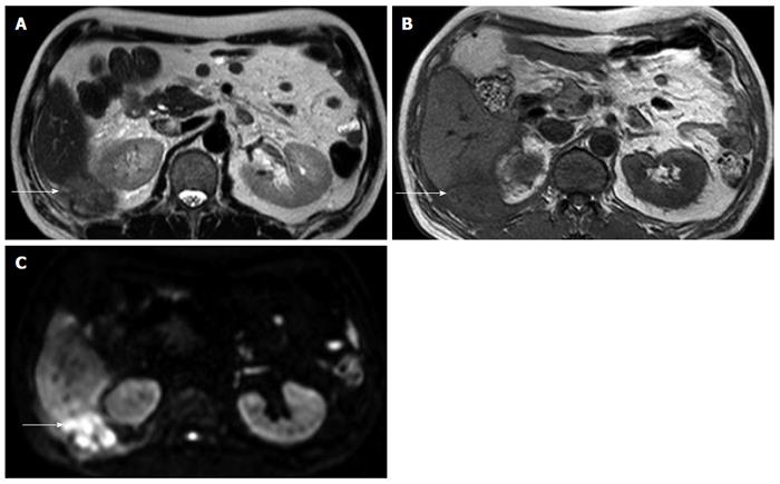
Figure 2 Magnetic resonance imaging showed ill-defined mass at right subhepatic space (arrows).
A: Subtle high signal intensity in T2 weighted image; B: Low signal intensity in T1 weighted image; C: High signal intensity in diffusion weighted image.
- Citation: Kim BS, Joo SH, Kim HC. Spilled gallstones mimicking a retroperitoneal sarcoma following laparoscopic cholecystectomy. World J Gastroenterol 2016; 22(17): 4421-4426
- URL: https://www.wjgnet.com/1007-9327/full/v22/i17/4421.htm
- DOI: https://dx.doi.org/10.3748/wjg.v22.i17.4421


