Copyright
©The Author(s) 2015.
World J Gastroenterol. Apr 7, 2015; 21(13): 4082-4088
Published online Apr 7, 2015. doi: 10.3748/wjg.v21.i13.4082
Published online Apr 7, 2015. doi: 10.3748/wjg.v21.i13.4082
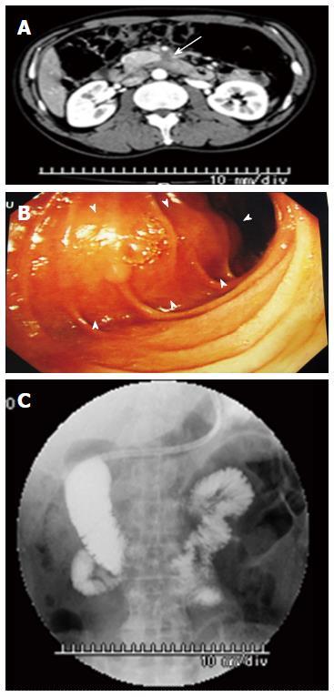
Figure 1 Contrast-enhanced computed tomography, duodenofiberoscopy, and hypotonic duodenography.
A: Abdominal CT scan shows a poorly enhanced mass around the third portion of the duodenum (arrow); B: Duodenofiberoscopic findings reveal submucosal tumors at the third portion of the duodenum (arrowheads); C: Hypotonic duodenography reveals stenosis at the third portion of the duodenum. CT: Computed tomography.
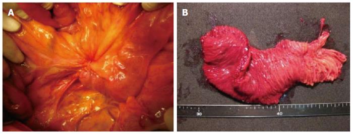
Figure 2 Intraoperative findings.
A: During surgery, the tumor with the umbilication was located in front of the Treitz ligament; B: Examination of the resected specimen revealed a normal duodenal surface.
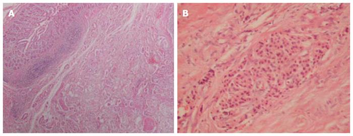
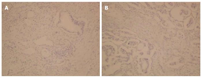
Figure 3 Histological examinations.
A: The resected part of the duodenum shows a moderately differentiated tubular adenocarcinoma [Hematoxylin and eosin (HE), × 40]; B: Only the islet cells are present within and near the tumor (HE, × 200). HE: Hematoxylin and eosin.
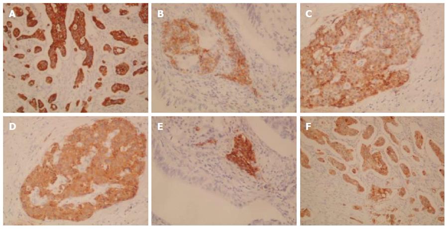
Figure 4 Immunohistochemical staining: positive staining.
A: Cytokeratin (CK) 7 (× 100); B: Neural cell adhesion molecule (CD56) (× 200); C: Chromogranin A (× 200); D: Synaptophysin (× 200); E: Insulin (× 200); F: Mucin 1 (MUC1).
Figure 5 Immunohistochemical staining: negative staining.
A: Cytokeratin (CK) 20 (× 100); B: Mucin 2 (MUC2) (× 200).
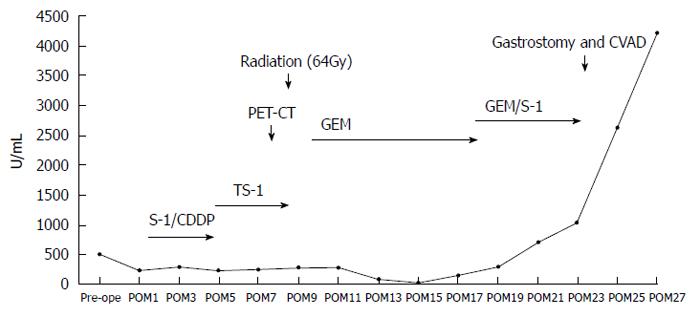
Figure 6 Chronological change of the serum level of CA19-9.
Pre-ope: Preoperation; POM: Post operative month; S-1: Tegafur-gimeracil-ditrydropyrimidine dehydrogenase; CDDP: Cisplatin; PET-CT: Positron emission and computed tomography; GEM: Gemcitabine; CVAD: Central venous access device.
- Citation: Fukino N, Oida T, Mimatsu K, Kuboi Y, Kida K. Adenocarcinoma arising from heterotopic pancreas at the third portion of the duodenum. World J Gastroenterol 2015; 21(13): 4082-4088
- URL: https://www.wjgnet.com/1007-9327/full/v21/i13/4082.htm
- DOI: https://dx.doi.org/10.3748/wjg.v21.i13.4082


