Copyright
©The Author(s) 2015.
World J Gastroenterol. Apr 7, 2015; 21(13): 4082-4088
Published online Apr 7, 2015. doi: 10.3748/wjg.v21.i13.4082
Published online Apr 7, 2015. doi: 10.3748/wjg.v21.i13.4082
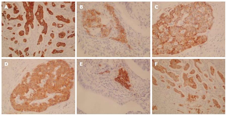
Figure 4 Immunohistochemical staining: positive staining.
A: Cytokeratin (CK) 7 (× 100); B: Neural cell adhesion molecule (CD56) (× 200); C: Chromogranin A (× 200); D: Synaptophysin (× 200); E: Insulin (× 200); F: Mucin 1 (MUC1).
- Citation: Fukino N, Oida T, Mimatsu K, Kuboi Y, Kida K. Adenocarcinoma arising from heterotopic pancreas at the third portion of the duodenum. World J Gastroenterol 2015; 21(13): 4082-4088
- URL: https://www.wjgnet.com/1007-9327/full/v21/i13/4082.htm
- DOI: https://dx.doi.org/10.3748/wjg.v21.i13.4082


