Copyright
©The Author(s) 2015.
World J Gastroenterol. Mar 21, 2015; 21(11): 3414-3419
Published online Mar 21, 2015. doi: 10.3748/wjg.v21.i11.3414
Published online Mar 21, 2015. doi: 10.3748/wjg.v21.i11.3414
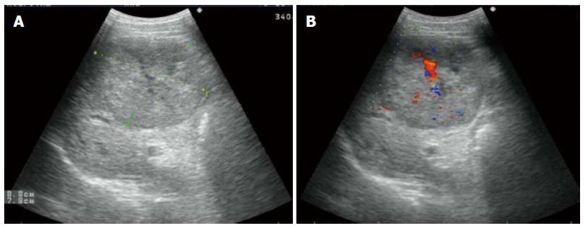
Figure 1 Tumor was hypoechoic on ultrasonography.
A: Measuring 8.8 cm × 7.8 cm well-defined hypoechoic mass; B: Color Doppler sonography showing a filiform vascular distribution pattern.
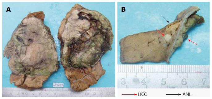
Figure 2 Macroscopical features of the tumors in this case.
A: The 10% neutral buffered formalin-fixed tumor occupied a large area of the right lobe. The encapsulated tumor was grayish-white and grayish-green. Scarring and broad fibrous septa was observed; B: Underneath the tumor capsule, an adjacent small yellowish soft area measuring 1.0 cm in diameter was observed, along with a small white nodule in it.
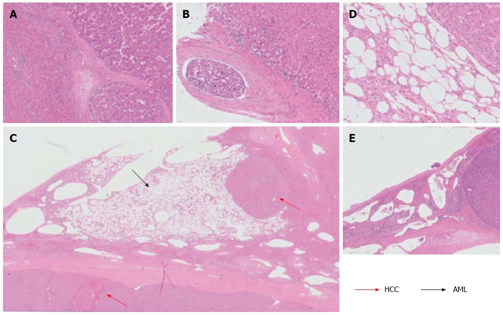
Figure 3 Histological features of the tumors in this case.
A: The HCC, showing a poor- to moderately-differentiation level, was demarcated from the surrounding liver tissue with a relatively clear boundary (HE staining, magnification × 50); B: A tumor emboli of HCC was found in a blood vessel located outside of the HCC mass (HE staining, magnification × 200); C and D: The AML component was composed of smooth muscle cells, adipose cells and blood vessels. The 0.3 cm white nodule in the middle of the AML was an HCC nodule with the same composition as the 9.0 cm HCC mass (C: HE staining, magnification × 10; D: HE staining, magnification × 200); E: The cavernous hemangioma areas were composed of large thin-walled vascular spaces, lined by a monolayer flat endothelial cells (HE staining, magnification × 50). HCC: Hepatocellular carcinoma; AML: Angiomyolipoma; HE: Hematoxylin and eosin.
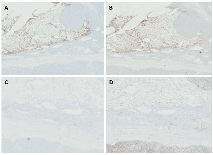
Figure 4 Immunohistochemical stainings.
A and B: The AML cells were positive for HMB45 (magnification × 10) and for A103 staining (magnification × 10); C and D: The AML cells were negative for Desmin (magnification × 20) and for CD34 staining (magnification × 20). AML: Angiomyolipoma.
- Citation: Ge XW, Zeng HY, Su-Jie A, Du M, Ji Y, Tan YS, Hou YY, Xu JF. Hepatocellular carcinoma with concomitant hepatic angiomyolipoma and cavernous hemangioma in one patient. World J Gastroenterol 2015; 21(11): 3414-3419
- URL: https://www.wjgnet.com/1007-9327/full/v21/i11/3414.htm
- DOI: https://dx.doi.org/10.3748/wjg.v21.i11.3414


