Copyright
©The Author(s) 2015.
World J Gastroenterol. Mar 21, 2015; 21(11): 3414-3419
Published online Mar 21, 2015. doi: 10.3748/wjg.v21.i11.3414
Published online Mar 21, 2015. doi: 10.3748/wjg.v21.i11.3414
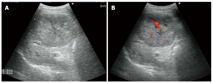
Figure 1 Tumor was hypoechoic on ultrasonography.
A: Measuring 8.8 cm × 7.8 cm well-defined hypoechoic mass; B: Color Doppler sonography showing a filiform vascular distribution pattern.
- Citation: Ge XW, Zeng HY, Su-Jie A, Du M, Ji Y, Tan YS, Hou YY, Xu JF. Hepatocellular carcinoma with concomitant hepatic angiomyolipoma and cavernous hemangioma in one patient. World J Gastroenterol 2015; 21(11): 3414-3419
- URL: https://www.wjgnet.com/1007-9327/full/v21/i11/3414.htm
- DOI: https://dx.doi.org/10.3748/wjg.v21.i11.3414


