Copyright
©2014 Baishideng Publishing Group Inc.
World J Gastroenterol. Nov 21, 2014; 20(43): 16372-16376
Published online Nov 21, 2014. doi: 10.3748/wjg.v20.i43.16372
Published online Nov 21, 2014. doi: 10.3748/wjg.v20.i43.16372
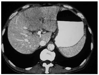
Figure 1 Computed tomography displaying a large low-density shadow in the left liver lobe.
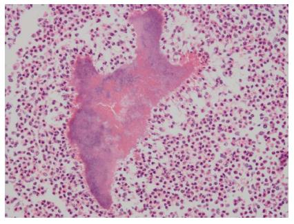
Figure 2 Sulphur particles surrounded by neutrophilic granulocyte infiltration (HE staining, × 100).
- Citation: Yang XX, Lin JM, Xu KJ, Wang SQ, Luo TT, Geng XX, Huang RG, Jiang N. Hepatic actinomycosis: Report of one case and analysis of 32 previously reported cases. World J Gastroenterol 2014; 20(43): 16372-16376
- URL: https://www.wjgnet.com/1007-9327/full/v20/i43/16372.htm
- DOI: https://dx.doi.org/10.3748/wjg.v20.i43.16372


