Copyright
©2014 Baishideng Publishing Group Inc.
World J Gastroenterol. Oct 28, 2014; 20(40): 14831-14840
Published online Oct 28, 2014. doi: 10.3748/wjg.v20.i40.14831
Published online Oct 28, 2014. doi: 10.3748/wjg.v20.i40.14831
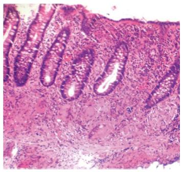
Figure 1 Histopathologic image.
Multiple mucosal and submucosal epithelioid cell granulomas with Langhan’s giant cells in a case of colonic tuberculosis.
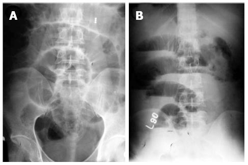
Figure 2 Plain X ray images.
Plain abdominal radiographs in supine (A) and erect (B) position showing dilated small bowel loops with air fluid levels in a patient who presented with subacute intestinal obstruction secondary to tubercular ileal stricture.
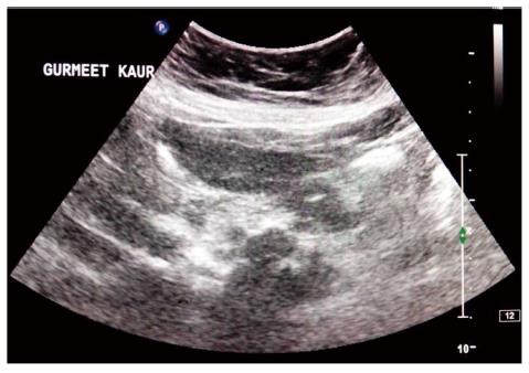
Figure 3 Ultrasound image.
Multiple enlarged conglomerate lymphnodes in retroperitoneum with hypoechoic centers due to caseation.
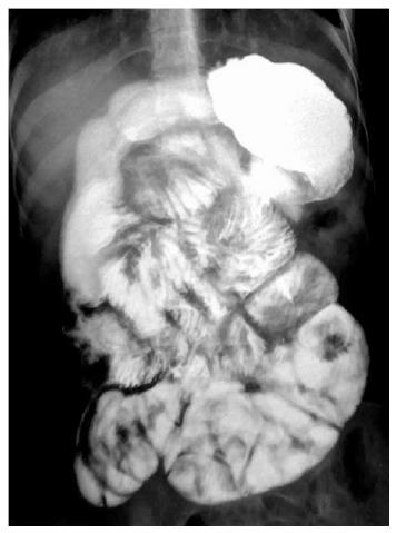
Figure 4 Barium meal follow through image.
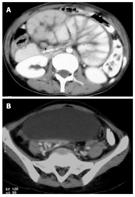
An adolescent female patient who was a case of tuberculosis with sclerosing encapsulating peritonitis (abdominal cocoon) showing clustering of small bowel loops in the centre of the abdomen which are constant in location.
Figure 5 Computed tomography images.
A: Axial images of the same patient showing small bowel loops congregated in the centre of the abdomen encased by a soft tissue density membrane. Enlarged discrete lymph nodes are also seen in the mesentery (arrow head); B: Loculated ascites is seen just below the level of conglomerate bowel loops.
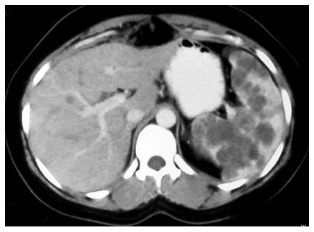
Figure 6 Computed tomography images.
Axial image of a young female, a case of tuberculosis showing few small ill-defined hypodense lesions in liver and multiple small hypodense lesions studded in the parenchyma of the spleen.
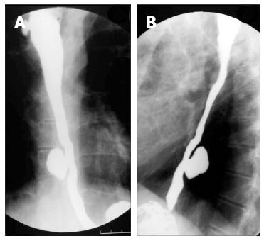
Figure 7 Barium swallow image.
An old treated case of pulmonary tuberculosis showing traction diverticulum in right lateral wall of upper thoracic esophagus (A). There is also a long segment smooth stricture in the distal thoracic esophagus with pulsion diverticulum in the right lateral wall of distal thoracic esophagus (A, B).
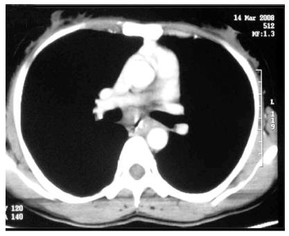
Figure 8 Computed tomography images.
Contrast enhanced computed tomography with oral contrast of another case of esophageal tuberculosis showing circumferential mural thickening in the mid thoracic esophagus. Histopathological examination of biopsies from the involved segments showed epithelioid granulomas.
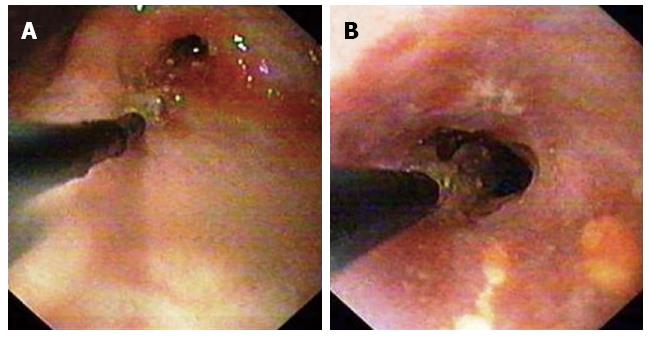
Figure 9 Endoscopic images.
Gastric tuberculosis with gastric outlet obstruction - before (A) and after (B) balloon dilatation.
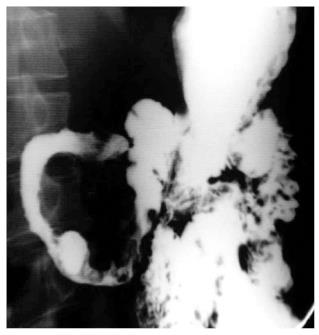
Figure 10 Barium meal follow through image.
Long segment circumferential narrowing in first and second part of duodenum.
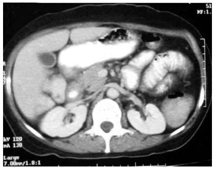
Figure 11 Computed tomography images.
Same patient showing smooth circumferential mural thickening involving the duodenum (D1 and D2). Biopsy showed granulomatous inflammation consistent with tuberculosis. Few enlarged subcentimetric lymphnodes are also seen in the mesentery.
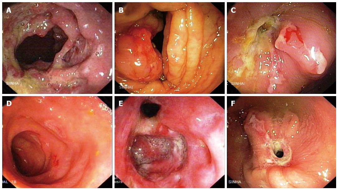
Figure 12 Endoscopic images.
A: Ulcerative form of ileocaecal (IC) tuberculosis - multiple ulceration on IC valve, caecum and ascending colon with nodularity in intervening area and some mucosal bridges; B: Hypertrophic form of ileocaecal tuberculosis - mass like lesion on IC valve with ulceration on surface; C: Ileocaecal tuberculosis - contracted caecum, narrowed and deformed IC valve and multiple ulceration on IC valve, caecum and ascending colon; D: Superficial ulcers in terminal ileum; E: Gaping IC valve with multiple ulcers on IC valve, caecum and ascending colon; F: Terminal ileal stricture with multiple ulcers on ileocaecal valve and contracted caecum.
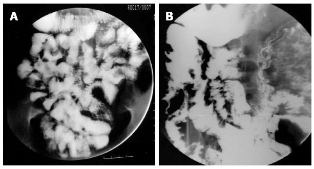
Figure 13 Barium meal follow through images.
A: A young patient who was a known case of peritoneal tuberculosis (TB) showing matting of small bowel loops; B: Luminal narrowing with ulcerations involving the terminal ileum and ileocaecal junction with contracted cecum- characteristic of ileocecal TB.
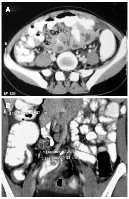
Figure 14 Computed tomography images.
A: Multiple conglomerate necrotic lymphnodes in the mesentery with contiguous involvement of the adjoining small bowel. Also noted is omental thickening and stranding; B: Coronal reformatted images of another patient showing smooth mural thickening in the terminal ileum and ileocecal junction. Large necrotic lymphnodes in the right iliac fossa are also seen.
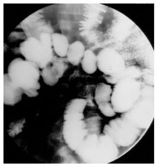
Figure 15 Barium enteroclysis image.
Multiple concentric short segment strictures in the ileum in a case of gastrointestinal tuberculosis.
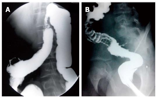
Figure 16 Barium enema images.
A: Tuberculous colitis presenting with concentric stricture in the distal transverse colon and splenic flexure. A fistulous tract is seen arising from hepatic flexure of colon with ulcerations in adjacent colon; B: Another patient with tuberculous colitis presenting with mucosal irregularity, ulcers in the sigmoid colon complicated by fistula formation in the distal sigmoid colon.
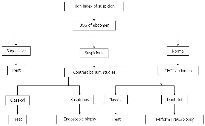
Figure 17 Management algorithm for abdominal tuberculosis.
Reproduced from Sood et al[41]. USG: Ultrasonography; CECT: Contrast enhanced computed tomography.
- Citation: Debi U, Ravisankar V, Prasad KK, Sinha SK, Sharma AK. Abdominal tuberculosis of the gastrointestinal tract: Revisited. World J Gastroenterol 2014; 20(40): 14831-14840
- URL: https://www.wjgnet.com/1007-9327/full/v20/i40/14831.htm
- DOI: https://dx.doi.org/10.3748/wjg.v20.i40.14831


