Copyright
©2014 Baishideng Publishing Group Inc.
World J Gastroenterol. Oct 28, 2014; 20(40): 14831-14840
Published online Oct 28, 2014. doi: 10.3748/wjg.v20.i40.14831
Published online Oct 28, 2014. doi: 10.3748/wjg.v20.i40.14831
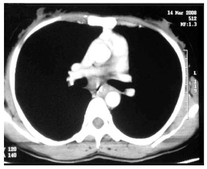
Figure 8 Computed tomography images.
Contrast enhanced computed tomography with oral contrast of another case of esophageal tuberculosis showing circumferential mural thickening in the mid thoracic esophagus. Histopathological examination of biopsies from the involved segments showed epithelioid granulomas.
- Citation: Debi U, Ravisankar V, Prasad KK, Sinha SK, Sharma AK. Abdominal tuberculosis of the gastrointestinal tract: Revisited. World J Gastroenterol 2014; 20(40): 14831-14840
- URL: https://www.wjgnet.com/1007-9327/full/v20/i40/14831.htm
- DOI: https://dx.doi.org/10.3748/wjg.v20.i40.14831


