
















©2014 Baishideng Publishing Group Inc.
World J Gastroenterol. Oct 28, 2014; 20(40): 14831-14840
Published online Oct 28, 2014. doi: 10.3748/wjg.v20.i40.14831
Published online Oct 28, 2014. doi: 10.3748/wjg.v20.i40.14831
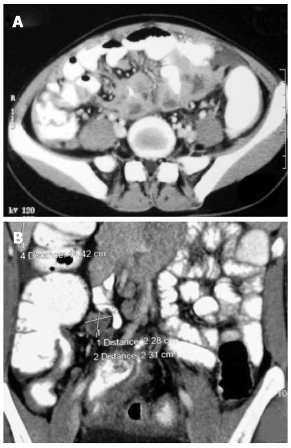
Figure 14 Computed tomography images.
A: Multiple conglomerate necrotic lymphnodes in the mesentery with contiguous involvement of the adjoining small bowel. Also noted is omental thickening and stranding; B: Coronal reformatted images of another patient showing smooth mural thickening in the terminal ileum and ileocecal junction. Large necrotic lymphnodes in the right iliac fossa are also seen.
- Citation: Debi U, Ravisankar V, Prasad KK, Sinha SK, Sharma AK. Abdominal tuberculosis of the gastrointestinal tract: Revisited. World J Gastroenterol 2014; 20(40): 14831-14840
- URL: https://www.wjgnet.com/1007-9327/full/v20/i40/14831.htm
- DOI: https://dx.doi.org/10.3748/wjg.v20.i40.14831