Copyright
©The Author(s) 1996.
World J Gastroenterol. Dec 15, 1996; 2(4): 238-240
Published online Dec 15, 1996. doi: 10.3748/wjg.v2.i4.238
Published online Dec 15, 1996. doi: 10.3748/wjg.v2.i4.238
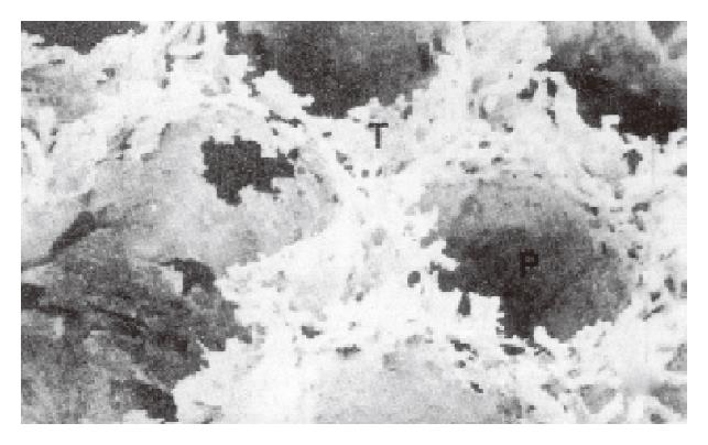
Figure 1 A scanning electron micrograph of lymphatic corrosion cast.
The lymphatic capillary plexus (T) in the TDA is connected with a perifollicular lymphatic sinus (P). (SEM, × 40)
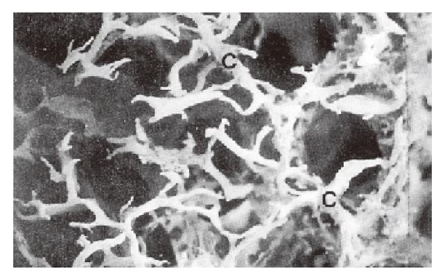
Figure 2 A scanning electron micrograph of lymphatic corrosion casts.
A well-developed network of lymphatic capillaries (C) exists in the superficial layer of the appendix mucosa. (SEM, ×30)
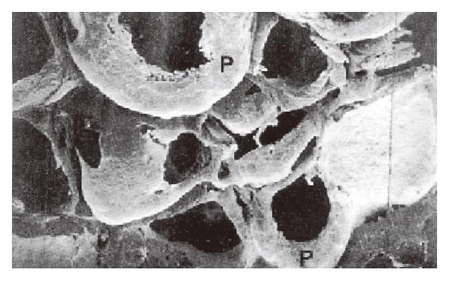
Figure 3 A scanning electron micrograph of lymphatic corrosion cast, displaying the bottoms of the perifollicular lymphatic sinuses (P).
(SEM, × 40)
- Citation: Tang FC, Zhang YF, Xu YD, Zhong SQ, Wang XP, Wang YX. Scanning electron microscopic study of lymphatic corrosion casts in the rabbit appendix. World J Gastroenterol 1996; 2(4): 238-240
- URL: https://www.wjgnet.com/1007-9327/full/v2/i4/238.htm
- DOI: https://dx.doi.org/10.3748/wjg.v2.i4.238


