Copyright
©2013 Baishideng Publishing Group Co.
World J Gastroenterol. Feb 28, 2013; 19(8): 1330-1332
Published online Feb 28, 2013. doi: 10.3748/wjg.v19.i8.1330
Published online Feb 28, 2013. doi: 10.3748/wjg.v19.i8.1330
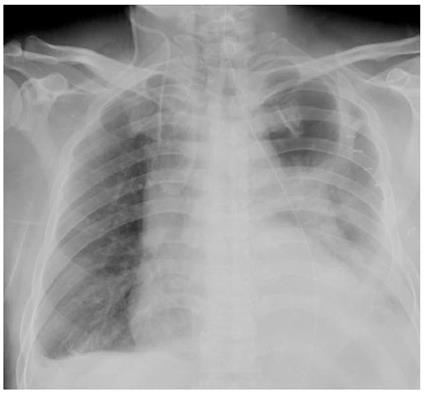
Figure 1 Postoperative chest X-ray.
A large consolidation shadow of the left lung, low density radiolucent areas on the left upper lobe and high density fragment images on the right lung are shown. Both costophrenic angles were obscure.
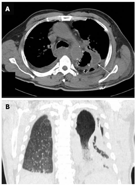
Figure 2 Computed tomography scan.
A: Axial computed tomography (CT) imaging of the chest: The CT scan showed a remarkably distended thoracic stomach and a locally thickened gastric wall (arrow); B: Coronal CT scan of the chest: Imaging revealed a thoracic stomach with a large consolidation shadow and in air bronchograms of the left lung, pleural and pericardial effusions were also suggested.
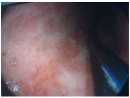
Figure 3 Endoscopic examination of the postoperative stomach.
The gastric mucosa was red and white in color, significantly edematous, and scattered erosions covered by purulent exudate were observed. No anastomotic leak was noted.
- Citation: Fan JQ, Liu DR, Li C, Chen G. Phlegmonous gastritis after esophagectomy: A case report. World J Gastroenterol 2013; 19(8): 1330-1332
- URL: https://www.wjgnet.com/1007-9327/full/v19/i8/1330.htm
- DOI: https://dx.doi.org/10.3748/wjg.v19.i8.1330


