Copyright
©2013 Baishideng Publishing Group Co.
World J Gastroenterol. Feb 28, 2013; 19(8): 1330-1332
Published online Feb 28, 2013. doi: 10.3748/wjg.v19.i8.1330
Published online Feb 28, 2013. doi: 10.3748/wjg.v19.i8.1330
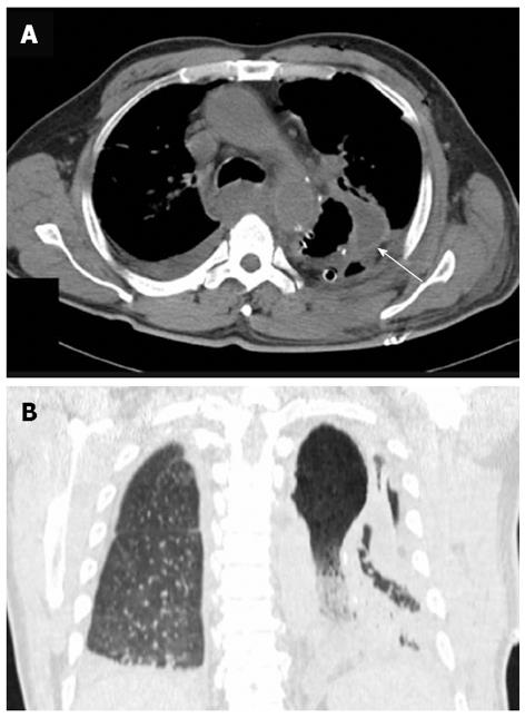
Figure 2 Computed tomography scan.
A: Axial computed tomography (CT) imaging of the chest: The CT scan showed a remarkably distended thoracic stomach and a locally thickened gastric wall (arrow); B: Coronal CT scan of the chest: Imaging revealed a thoracic stomach with a large consolidation shadow and in air bronchograms of the left lung, pleural and pericardial effusions were also suggested.
- Citation: Fan JQ, Liu DR, Li C, Chen G. Phlegmonous gastritis after esophagectomy: A case report. World J Gastroenterol 2013; 19(8): 1330-1332
- URL: https://www.wjgnet.com/1007-9327/full/v19/i8/1330.htm
- DOI: https://dx.doi.org/10.3748/wjg.v19.i8.1330


