Copyright
©2013 Baishideng Publishing Group Co.
World J Gastroenterol. Feb 28, 2013; 19(8): 1330-1332
Published online Feb 28, 2013. doi: 10.3748/wjg.v19.i8.1330
Published online Feb 28, 2013. doi: 10.3748/wjg.v19.i8.1330
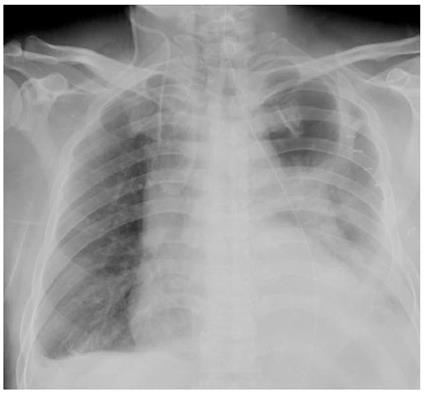
Figure 1 Postoperative chest X-ray.
A large consolidation shadow of the left lung, low density radiolucent areas on the left upper lobe and high density fragment images on the right lung are shown. Both costophrenic angles were obscure.
- Citation: Fan JQ, Liu DR, Li C, Chen G. Phlegmonous gastritis after esophagectomy: A case report. World J Gastroenterol 2013; 19(8): 1330-1332
- URL: https://www.wjgnet.com/1007-9327/full/v19/i8/1330.htm
- DOI: https://dx.doi.org/10.3748/wjg.v19.i8.1330


